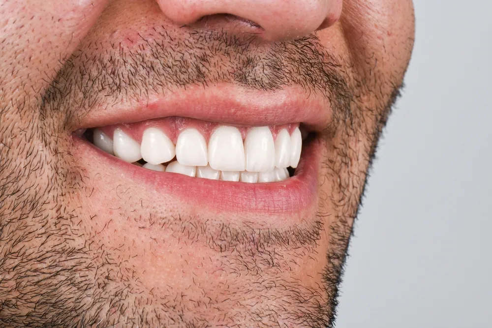
Yes, E.max veneers can be safe when dentists select appropriate cases and follow conservative preparation and bonding protocols. The ceramic itself shows strong biocompatibility and mechanical performance. However, safety depends on multiple factors beyond the material alone.
E.max veneers represent one of the most popular choices in modern esthetic dentistry. These thin restorations generally come from lithium disilicate glass-ceramic. Dentists place them on the front surfaces of teeth to improve color, shape, and alignment. Over the past decade, E.max has gained widespread use because it combines strength with natural appearance. But patients still ask one central question: Does this material pose real risks? The answer requires careful examination. Material safety differs from procedural safety. The ceramic itself rarely causes biological harm. Most complications arise from treatment technique, patient factors, and long-term maintenance. This article examines the biological, mechanical, functional, and long-term safety of E.max veneers. Current evidence supports predictable clinical outcomes when clinicians follow proper protocols. The major safety factors include tooth preservation, enamel preparation, adhesive bonding, occlusal forces, fracture resistance, debonding risk, gingival health, oral hygiene, and long-term maintenance (Beier et al. 2012, 173).
What Are E.max Veneers Made Of?
E.max veneers consist of lithium disilicate glass-ceramic. This material combines high strength with excellent translucency. Manufacturers produce these veneers through heat-pressing or CAD/CAM milling.
What Is Lithium Disilicate Ceramic?
Lithium disilicate is a glass-ceramic material. It contains lithium dioxide and silicon dioxide crystals. This composition gives it both strength and a tooth-like appearance.
Lithium disilicate belongs to the glass-ceramic family. It contains fine lithium disilicate crystals within a glassy matrix. This structure gives the material unique properties. It offers high flexural strength. It resists fracture under normal chewing forces. It also transmits light in a way that mimics natural enamel. Dentists value this combination. Older ceramic materials often forced clinicians to choose between strength and beauty. Lithium disilicate provides both. The material scores approximately 360 to 400 MPa in flexural strength tests. This measurement exceeds the values for traditional feldspathic porcelain. Yet lithium disilicate maintains better translucency than zirconia. This balance makes it ideal for anterior veneers. The ceramic also shows excellent biocompatibility. Oral tissues tolerate it well. Researchers have documented its safe use in thousands of clinical cases (Layton and Walton 2012, 1045).
How Are E.max Veneers Manufactured?
Dental labs manufacture E.max veneers through heat-pressing or CAD/CAM milling. Both methods produce reliable restorations when technicians follow proper protocols.
Manufacturers use two main techniques to create E.max veneers. The first method involves heat-pressing. Technicians wax a veneer pattern and then press molten lithium disilicate ceramic into a mold. The second method uses CAD/CAM technology. A milling machine carves the veneer from a pre-crystallized block. The lab then heat-treats the restoration to achieve final strength. Some veneers use a monolithic approach. This means the entire veneer comes from one uniform material. Other veneers use a layered approach. Technicians add veneering porcelain over a lithium disilicate core. The manufacturing method influences the final result. Milled veneers often show excellent marginal adaptation. Pressed veneers can provide superior esthetics in some cases. Recent systematic reviews report satisfactory clinical outcomes for both techniques. A 2025 systematic review of milled versus heat-pressed lithium disilicate veneers found satisfactory outcomes with both fabrication methods. However, the review also noted low or very low certainty for several outcomes because of methodological limitations in existing studies (Etemadi Saberi et al. 2021, 647).
Are E.max Veneers Biocompatible?
Yes, lithium disilicate shows excellent biocompatibility. Oral tissues tolerate this ceramic well. Most biological complications stem from restoration fit and patient hygiene rather than the material itself.
Is Lithium Disilicate Safe for Oral Tissues?
Lithium disilicate ceramic proves safe for oral tissues. It does not release harmful substances. It does not irritate the gums or soft tissues when dentists place it properly.
Biocompatibility means a material can coexist with living tissues without causing harm. Dental materials must meet strict biocompatibility standards. Lithium disilicate has undergone extensive testing. Research shows that this ceramic does not trigger toxic responses in oral tissues. It does not dissolve or leach chemicals into saliva. It does not alter the oral microbiome in harmful ways. The material remains chemically stable in the wet oral environment. However, the ceramic composition alone does not guarantee biological success. The way the dentist designs and places the veneer matters just as much. Poor margins can trap plaque. Overcontoured veneers can impinge on gum tissue. Inadequate polishing can harbor bacteria. These factors cause biological complications. But the material itself rarely triggers adverse tissue reactions (Peumans et al. 2000, 165).
Can E.max Veneers Cause Allergic Reactions?
True allergic reactions to lithium disilicate ceramic are extremely rare. Most oral sensitivities relate to other factors like bonding agents or cements.
True material hypersensitivity to dental ceramics remains uncommon. Lithium disilicate contains inorganic compounds. These compounds rarely provoke immune responses. True allergies typically involve proteins or organic compounds. Some patients report oral sensitivity after veneer placement. But this sensitivity usually stems from other causes. The bonding resin or cement might irritate some individuals. The tooth preparation itself might cause temporary nerve irritation. Patients should always disclose previous reactions to dental materials. This disclosure helps the dentist select appropriate bonding systems. Individualized dental assessment remains essential. No dentist should assume every patient has identical material tolerance. A thorough medical history prevents most complications (Stokes and Palmer 2005, 427).
Can E.max Veneers Irritate the Gums?
Well-made E.max veneers do not irritate gums. Poorly adapted margins, overcontouring, or inadequate polishing can cause gingival inflammation.
Gingival health depends on multiple factors. Accurate margins play a critical role. The veneer edge must fit flush against the tooth. Proper contour matters too. The veneer should follow the natural tooth shape. Smooth surfaces resist plaque accumulation. Patients must maintain adequate oral hygiene. The gums should be healthy before treatment begins. Poorly adapted restorations can trap bacteria. This bacterial accumulation leads to gingival inflammation. But this problem stems from restoration design and execution. The lithium disilicate material itself does not cause gum irritation. A well-designed E.max veneer with proper margins and smooth surfaces supports periodontal health (Gurel 2003, 19).
Does Getting E.max Veneers Damage Natural Teeth?
E.max veneer preparation removes some tooth structure. Dentists use conservative techniques to preserve enamel. This preparation is generally irreversible.
How Much Tooth Structure Is Removed for E.max Veneers?
Preparation depth varies by case. Most dentists remove 0.3 to 0.7 millimeters of enamel. Some cases require slightly more reduction.
Conservative veneer preparation forms the foundation of safe treatment. Dentists remove a thin layer of enamel to create space for the ceramic. The exact depth depends on several factors. The existing tooth color matters. Darker teeth may need more reduction to allow proper masking. The desired final shade influences preparation depth. Tooth position affects the plan. Central incisors often need different preparation than canines. The veneer thickness must accommodate the ceramic strength. Occlusion guides the design. The dentist must consider how the upper and lower teeth meet. Restoration design also determines preparation extent. Some cases need minimal reduction. Other cases require more extensive preparation. But "minimal preparation" does not mean "no preparation" in every case. Patients should understand that some enamel removal always occurs (Edelhoff and Beuer 2002, 503).
Why Is Enamel Preservation Important for Veneer Safety?
Enamel preservation supports strong adhesive bonding. It reduces unnecessary dentin exposure. It helps maintain tooth vitality and structural integrity.
Enamel bonding drives veneer success. The adhesive system works best on enamel. Enamel provides a hard, mineralized surface. This surface allows resin cement to form strong micromechanical retention. Maintaining enamel supports predictable bonding. It reduces the risk of debonding. It also reduces unnecessary dentin exposure. Dentin contains tubules that lead to the tooth nerve. Excessive dentin exposure can increase sensitivity. It can also raise the risk of pulpal inflammation. Dentists should preserve as much healthy enamel as clinically possible. This preservation supports both immediate bonding and long-term tooth health (Magne and Belser 2004, 9).
Are E.max Veneers Reversible?
No, conventional E.max veneer treatment is generally irreversible. The dentist removes enamel, and enamel does not regenerate.
Patients often ask whether they can reverse veneer treatment. The honest answer is no. Once the dentist removes enamel, the body cannot replace it. Some marketing materials suggest "no-prep" or "minimal-prep" veneers as reversible options. But even minimal preparation removes some enamel. True reversible treatment would require zero tooth reduction. In most cases, some reduction proves necessary for proper fit and esthetics. Patients should understand this before starting treatment. They should also understand that veneers require replacement over time. No veneer lasts forever. Future replacement procedures may require additional tooth modification. Informed consent must include this long-term perspective (Calamia and Calamia 2007, 405).
Are E.max Veneers Strong Enough for Daily Use?
Yes, E.max veneers withstand normal chewing forces. Their strength comes from lithium disilicate crystals. However, thickness, bonding, and tooth support all influence performance.
How Strong Are E.max Veneers?
Laboratory tests show flexural strength around 360 to 400 MPa. This exceeds traditional feldspathic porcelain. Real-world performance depends on proper design and bonding.
Lithium disilicate offers impressive mechanical properties. Laboratory tests measure its flexural strength at approximately 360 to 400 MPa. This value exceeds the strength of traditional feldspathic porcelain by a significant margin. But laboratory strength does not automatically predict clinical longevity. Several factors influence real-world performance. Veneer thickness matters. Thinner veneers withstand less force than thicker ones. Veneer geometry influences stress distribution. Sharp angles concentrate force. Rounded designs distribute force better. The bonding quality affects strength. A well-bonded veneer receives support from the underlying tooth. The tooth itself must be healthy. A compromised tooth cannot support the veneer properly. So while the material shows excellent inherent strength, the overall restoration strength depends on the entire system (Guess et al. 2011, 335).
Are E.max Veneers Resistant to Chipping and Fracture?
E.max veneers resist chipping better than traditional porcelain. But no ceramic veneer is completely fracture-proof. Excessive force can still cause damage.
E.max veneers demonstrate good resistance to chipping and cracking. The lithium disilicate crystals interrupt crack propagation. This mechanism prevents small cracks from growing into large fractures. However, no ceramic restoration is indestructible. Unfavorable occlusion increases mechanical risk. If the veneer takes excessive biting force, it can fracture. Parafunctional habits like nail-biting or ice-chewing raise the risk. Poor veneer design can create weak points. A veneer that is too thin in one area may fracture there. Dentists must evaluate occlusal forces before placing veneers. They must design the restoration to handle expected functional loads. Patients must avoid using their teeth as tools. Realistic expectations remain important. E.max provides excellent fracture resistance, but it is not invincible (Fradeani and Barducci 2018, 170).
Can People With Bruxism Safely Have E.max Veneers?
People with bruxism can receive E.max veneers, but they need careful evaluation. Night guards and occlusal management become essential.
Bruxism requires special consideration. This condition involves grinding or clenching teeth, usually during sleep. These habits generate excessive forces. Such forces can overload ceramic veneers. Dentists must evaluate bruxism before recommending E.max veneers. They should assess the severity of the habit. They should examine the current tooth wear patterns. Bite stabilization may be necessary. A protective night guard becomes essential for most bruxism patients. This guard absorbs forces and protects the veneers. The restoration design may need modification. The dentist might increase veneer thickness in high-stress areas. They might adjust the occlusion to reduce point contacts. In some severe cases, the dentist may recommend an alternative material. Zirconia can offer greater fracture resistance for patients with extreme bruxism. Each case requires individualized assessment (Layton and Walton 2012, 1048).
How Long Do E.max Veneers Safely Last?
Clinical studies report high survival rates. Many E.max veneers function well for over 10 years. Proper care extends their service life.
What Does the Research Say About E.max Veneer Survival?
Research shows pooled survival rates around 96.8% at approximately 10.4 years. Individual outcomes vary by case selection and technique.
Clinical survival provides the most meaningful measure of veneer success. Manufacturer claims mean little without long-term patient data. Fortunately, researchers have tracked E.max veneer performance for many years. A recent systematic review and meta-analysis reported a pooled long-term survival rate of approximately 96.8% for lithium disilicate laminate veneers. The mean observation period in this analysis was 10.4 years. This data comes from multiple clinical studies. However, complication and survival outcomes vary according to study design and clinical circumstances. Some studies report even higher survival rates. Others show slightly lower figures. The variation reflects differences in patient selection, preparation protocols, and follow-up periods. Overall, the evidence strongly supports E.max as a reliable long-term restoration (Etemadi Saberi et al. 2021, 648).
What Factors Affect the Longevity of E.max Veneers?
Many factors influence veneer longevity. These include bonding quality, preparation design, veneer thickness, occlusion, bruxism, oral hygiene, diet, maintenance, lab quality, and clinician experience.
Multiple variables determine how long E.max veneers last. The quality of enamel bonding tops the list. Poor bonding leads to early failure. Tooth preparation design matters. Overly aggressive preparation weakens the tooth. Conservative preparation preserves strength. Veneer thickness affects durability. Very thin veneers may lack adequate strength. Occlusion plays a major role. Unbalanced bites concentrate destructive forces. Bruxism and clenching accelerate wear and fracture risk. Oral hygiene prevents decay at the margins. Dietary habits influence outcomes. Excessive consumption of hard foods raises fracture risk. Professional maintenance catches problems early. The quality of laboratory fabrication affects fit and strength. Finally, dentist and ceramist experience directly impacts success rates. Experienced teams achieve better outcomes (Beier et al. 2012, 175).
Do E.max Veneers Need to Be Replaced?
Yes, veneers require replacement eventually. They are not lifetime restorations. Replacement timing varies by patient and circumstance.
Veneers do not last forever. Every restoration has a finite lifespan. Several events can trigger replacement. Fracture obviously requires immediate replacement. Debonding may allow re-cementation, but often necessitates a new veneer. Wear can alter the surface texture over time. Marginal problems like decay or leakage require intervention. Esthetic changes may prompt replacement. The underlying tooth may darken. The cement may shift in color. Surrounding teeth may change shade. Patients should not expect a single universal replacement interval. Clinical longevity varies substantially between patients. Some veneers last 20 years or more. Others need replacement within 5 to 7 years. Regular dental checkups help monitor veneer condition. Early intervention prevents more extensive problems (Sasse and Thompson 2019, 790).
Can E.max Veneers Cause Tooth Sensitivity?
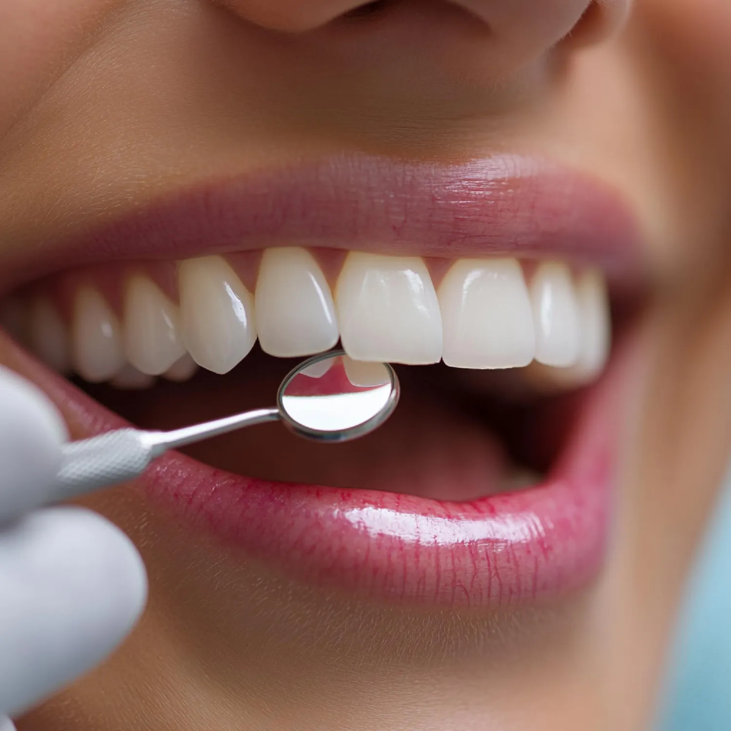
Some patients experience temporary sensitivity after veneer placement. This sensitivity usually resolves within days or weeks. Persistent sensitivity requires dental evaluation.
Why Can Sensitivity Occur After Veneer Placement?
Sensitivity can result from enamel reduction, dentin exposure, the cementation process, occlusal trauma, or pre-existing nerve irritation.
Temporary postoperative sensitivity is common. Several factors contribute to this sensation. Enamel reduction removes the tooth's natural insulating layer. This exposes the underlying dentin. Dentin contains microscopic tubules. These tubules connect to the tooth nerve. External stimuli can travel through these tubules. The cementation procedure itself may irritate the nerve. Acid etching and bonding agents can cause temporary pulpal inflammation. Occlusal trauma raises another concern. If the veneer sits too high, the tooth takes excessive biting force. This force can inflame the nerve. Pre-existing pulpal sensitivity may also surface after treatment. A tooth with a slightly inflamed nerve before preparation may become more sensitive afterward. Most cases resolve as the tooth heals. Anti-sensitivity toothpaste often helps during this period (Gurel 2003, 21).
Is Persistent Sensitivity Normal?
No, persistent sensitivity is not normal. Sensitivity lasting more than a few weeks warrants professional evaluation.
Patients should distinguish expected short-term sensitivity from persistent symptoms. Mild sensitivity to cold or air for a few days is normal. Sensitivity that lasts weeks or months is not. This persistent discomfort signals an underlying problem. The nerve may have sustained excessive trauma during preparation. The dentist may have removed too much tooth structure. Occlusal interference may overload the tooth. Leakage or bonding problems may allow fluid movement. In rare cases, the nerve may be irreversibly inflamed. This condition may require root canal treatment. Patients should report persistent sensitivity to their dentist immediately. Early diagnosis prevents more serious complications. The dentist can adjust the bite, replace the veneer, or recommend appropriate treatment (Edelhoff and Beuer 2002, 506).
Are E.max Veneers Safe for the Gums and Periodontal Health?
Yes, E.max veneers support gum health when dentists place them properly. Margin placement, contour, and surface finish matter more than the ceramic itself.
How Does Veneer Placement Affect Gingival Health?
Properly placed veneers do not harm gums. Supragingival margins and biologically appropriate contours protect periodontal tissues.
Gingival health depends heavily on restoration design. Dentists should place margins supragingivally when possible. This means the veneer edge sits above the gumline. Supragingival margins allow patients to clean the area easily. Biologically appropriate margins follow the natural tooth contour. The veneer should not invade the biological width. This term refers to the zone of gum attachment. Violating this space triggers inflammation. Plaque retention also threatens gum health. Overcontoured veneers create ledges where plaque accumulates. Well-contoured veneers with smooth surfaces resist plaque buildup. Periodontal health should be assessed before cosmetic treatment. Active gum disease must be treated first. Healthy gums provide the best foundation for veneers. A comprehensive periodontal evaluation protects long-term outcomes (Peumans et al. 2000, 168).
Can Poorly Fitted E.max Veneers Cause Gum Problems?
Yes, poorly fitted veneers can cause gum inflammation. Overcontouring, open margins, cement remnants, and rough surfaces all contribute to biological complications.
Poor restoration quality creates biological risks. Overcontouring pushes against the gum tissue. This pressure restricts blood flow. It also creates a plaque trap. Open or defective margins allow bacteria to penetrate. These bacteria cause chronic inflammation. Cement remnants left under the gumline act as irritants. Inadequate polishing leaves rough surfaces. Rough surfaces attract plaque more than smooth ones. Poor oral hygiene compounds these problems. Patients must brush and floss diligently. But even perfect hygiene cannot overcome a poorly designed veneer. The good news is that these complications relate to restoration design and execution. They do not stem from lithium disilicate itself. A well-made E.max veneer with excellent margins and polish supports healthy gums (Stokes and Palmer 2005, 428).
Are E.max Veneers Safer Than Traditional Porcelain Veneers?
E.max offers greater strength than traditional feldspathic porcelain. Both materials can be safe when dentists use them appropriately. The best choice depends on the specific case.
How Do E.max and Feldspathic Porcelain Differ?
E.max contains lithium disilicate crystals. Feldspathic porcelain is a traditional glass-based ceramic. E.max is stronger. Feldspathic porcelain offers slightly more translucency in some applications.
The two materials differ in composition and performance. Feldspathic porcelain has served dentistry for decades. It contains silica, alumina, and potassium oxide. It offers exceptional esthetics. It mimics natural enamel beautifully. However, it lacks strength. Its flexural strength ranges from 60 to 100 MPa. This low strength limits its use in high-stress situations. E.max contains lithium disilicate crystals. These crystals dramatically increase strength. E.max reaches 360 to 400 MPa. This strength allows thinner restorations. E.max veneers can be 0.3 to 0.5 millimeters thick. Feldspathic veneers often need more bulk for adequate strength. E.max also shows better fracture resistance. But feldspathic porcelain can achieve slightly more translucency in expert hands. Both materials require tooth preparation. The preparation depth depends on the desired outcome and existing tooth color. Material selection should match the clinical situation (Calamia and Calamia 2007, 402).
Is E.max More Durable Than Traditional Porcelain?
Yes, E.max generally outlasts feldspathic porcelain. Its higher strength reduces fracture and chipping. But patient factors still determine ultimate durability.
E.max holds a clear mechanical advantage over traditional feldspathic porcelain. The lithium disilicate structure resists crack propagation better. This means E.max veneers chip less often. They fracture less frequently under normal forces. Clinical studies support this laboratory data. E.max veneers show higher survival rates in long-term tracking. But framing one material as universally superior would be misleading. Patient-specific factors determine material selection. Some highly esthetic cases may still benefit from feldspathic porcelain. Skilled ceramists can create stunning results with either material. The patient's bite force, habits, and expectations all influence the choice. For most patients seeking anterior veneers, E.max provides an excellent balance of strength and beauty (Fradeani and Barducci 2018, 172).
Which Material Requires More Conservative Tooth Preparation?
Both materials allow conservative preparation. E.max often requires less reduction because its strength permits thinner designs.
Case-dependent factors determine preparation requirements. E.max strength allows very thin veneers. In some cases, dentists can place E.max veneers with minimal enamel reduction. Feldspathic porcelain needs slightly more bulk for strength. However, the existing tooth color influences preparation more than the material itself. A dark tooth requires more reduction regardless of the ceramic type. The dentist must create enough space to mask the underlying color. Enamel thickness also matters. Some patients have thick enamel. Others have thin enamel. The desired esthetic transformation guides the preparation plan. A minor shape change needs little reduction. A major color change needs more reduction. Neither material automatically guarantees a more conservative approach. The dentist must evaluate each tooth individually (Magne and Belser 2004, 11).
Are E.max Veneers Safer Than Zirconia Veneers?
E.max and zirconia both offer safety in appropriate cases. Zirconia provides greater strength. E.max provides superior esthetics for anterior teeth.
How Do E.max and Zirconia Compare in Strength?
Zirconia generally provides greater mechanical strength than E.max. Zirconia flexural strength can exceed 900 MPa. E.max reaches approximately 360 to 400 MPa.
Zirconium dioxide, commonly called zirconia, represents a different ceramic category. It belongs to the oxide ceramic family. Zirconia contains no glass phase. This composition gives it exceptional strength. Its flexural strength often exceeds 900 MPa. Some high-translucency zirconias offer slightly lower strength but still exceed E.max values. This superior strength makes zirconia ideal for posterior crowns and bridges. But higher strength does not automatically mean greater suitability for veneers. Anterior veneers require more than brute strength. They must transmit light naturally. They must blend with adjacent teeth. E.max outperforms most zirconias in translucency. The esthetic result often looks more lifelike with lithium disilicate. For anterior veneers, E.max frequently provides the better balance of strength and beauty (Zarone et al. 2011, 88).
Which Material Is More Esthetic?
E.max generally offers superior esthetics for anterior veneers. It transmits light more like natural enamel. Zirconia can appear slightly more opaque.
Esthetics drives most veneer decisions. Patients want restorations that look completely natural. E.max excels in this area. Its glass-ceramic structure allows excellent light transmission. Light enters the veneer and scatters naturally. This behavior mimics natural enamel. The result is a vibrant, lifelike appearance. Zirconia has improved dramatically in recent years. Newer high-translucency zirconias offer better esthetics than earlier versions. But most dentists still prefer E.max for highly esthetic anterior cases. E.max integrates better with surrounding natural teeth. It reflects light in a more enamel-like manner. Shade matching becomes easier. The margins blend more seamlessly. For patients seeking the most natural smile, E.max usually wins the esthetic comparison (Guess et al. 2011, 340).
When Might Zirconia Be Preferable?
Zirconia may work better for patients with severe bruxism, high functional loading, or specific masking requirements. Dentists must assess each case individually.
Zirconia shines in specific clinical situations. Patients with severe bruxism may benefit from its superior fracture resistance. High functional loading on anterior teeth may warrant the stronger material. Some cases require extensive masking of dark underlying tooth structure. Certain zirconia formulations provide better opacity for these situations. Cases requiring greater fracture resistance may also favor zirconia. But the decision always requires individualized clinical assessment. The dentist must weigh esthetics against mechanical demands. They must consider the patient's habits, bite forces, and expectations. For the average patient seeking anterior veneers, E.max remains the more popular choice. For the high-risk patient with extreme functional demands, zirconia may provide added security (Zarone et al. 2011, 90).
What Are the Main Risks and Complications of E.max Veneers?
The main risks include debonding, fracture, discoloration, and bite problems. Most complications relate to technique, design, or patient factors rather than the material itself.
Can E.max Veneers Debond?
Yes, debonding can occur. It usually results from bonding protocol errors, moisture contamination, or insufficient enamel.
Debonding means the veneer separates from the tooth. This is an adhesive failure. Several factors influence debonding risk. The bonding protocol must be precise. The dentist must isolate the tooth properly. Saliva or blood contamination weakens the bond. Enamel availability matters. Dentin bonding is less predictable than enamel bonding. If the dentist removes too much enamel, the veneer bonds to dentin instead. This increases debonding risk. Ceramic surface treatment also affects retention. The lab must etch the ceramic properly. Silane application improves the bond between ceramic and resin cement. Cement selection plays a role. Some cements work better with lithium disilicate than others. Proper polymerization ensures complete cement curing. Finally, the dentist must remove all excess cement. Leftover cement can cause inflammation and compromise the margin. Strict adherence to evidence-based adhesive protocols prevents most debonding failures (Aboushelib et al. 2006, 829).
Can E.max Veneers Crack or Fracture?
Yes, E.max veneers can crack or fracture. Excessive occlusal forces and inadequate design increase this risk.
Mechanical failure can occur even with strong materials. E.max resists fracture well, but it is not immune. Excessive occlusal forces create stress. These forces may come from normal chewing on unfavorably designed veneers. They may come from parafunctional habits like grinding. Inadequate design creates weak points. A veneer that is too thin in the incisal area may chip. A veneer with unsupported edges may crack. The tooth underneath must provide adequate support. A compromised tooth cannot reinforce the veneer properly. Patients must avoid biting hard objects. Pens, ice, and fingernails can fracture veneers. Night guards protect against nocturnal grinding forces. Proper design, bonding, and patient behavior together prevent most fractures (Layton and Walton 2012, 1047).
Can E.max Veneers Become Discolored?
The ceramic itself resists discoloration. But the cement, underlying tooth, or margins may change color over time.
Lithium disilicate ceramic shows excellent color stability. The material itself does not stain easily. It does not absorb pigments from coffee, tea, or wine. However, other components can change color. The resin cement may shift slightly over years. This is more common with older cement formulations. The underlying tooth structure may darken naturally. Natural teeth tend to yellow with age. A veneer cannot prevent this internal change. Margins may accumulate stain. Poor oral hygiene allows plaque and calculus buildup. These deposits appear yellow or brown. Surrounding restorations may also change color. An old filling next to a veneer may darken. This contrast makes the veneer appear mismatched. Regular professional cleanings help maintain color harmony. Good oral hygiene prevents surface staining at the margins (Peumans et al. 2000, 170).
Can E.max Veneers Cause Bite Problems?
Poorly adjusted veneers can disrupt occlusion. Dentists must check both static and dynamic contacts before finalizing the restoration.
Excessive veneer thickness can alter the bite. If the veneer sits too high, the tooth makes premature contact. This contact throws off the entire occlusal scheme. The patient may feel the tooth first when closing. This sensation indicates a high spot. Dynamic contacts also matter. These are the contacts that occur during chewing movements. The dentist must check lateral and protrusive movements. The veneer should not interfere with the natural glide of the jaw. Occlusal adjustment corrects minor interferences. The dentist uses articulating paper to mark contact points. They then polish these spots to achieve harmonious occlusion. Failure to address occlusal problems can lead to veneer fracture, tooth sensitivity, or jaw pain. Proper occlusal checking is a critical step in veneer delivery (Gurel 2003, 22).
Who Is a Good Candidate for E.max Veneers?
Good candidates have healthy teeth and gums, adequate enamel, realistic expectations, and stable occlusion. They must also commit to good oral hygiene.
What Dental Conditions Can E.max Veneers Correct?
E.max veneers can correct discoloration, minor shape problems, small gaps, chipped edges, mild wear, cosmetic asymmetry, and selected tooth-size discrepancies.
E.max veneers offer versatile cosmetic solutions. They mask discoloration that does not respond to bleaching. They correct minor shape abnormalities. A peg-shaped lateral incisor can gain normal proportions. They close small gaps between teeth. They restore chipped incisal edges. They cover mild wear from aging or erosion. They improve cosmetic asymmetry. They can address selected cases of tooth-size discrepancy. But veneers cannot fix every dental problem. They do not correct major misalignment. They do not treat active decay. They do not replace missing teeth. They work best for patients with fundamentally healthy dentition who seek esthetic enhancement (Fradeani and Barducci 2018, 175).
Who May Not Be a Good Candidate?
Poor candidates include patients with active gum disease, extensive decay, insufficient enamel, unmanaged bruxism, unstable bites, poor oral hygiene, or unrealistic expectations.
Not everyone qualifies for veneer treatment. Active untreated periodontal disease disqualifies a patient. The gums must be healthy first. Extensive untreated caries require restorative treatment, not veneers. Insufficient enamel compromises bonding. The veneer needs enamel for predictable retention. Severe bruxism without management raises fracture risk. An unstable occlusion needs correction before cosmetic work. Poor oral hygiene leads to decay and gum problems around veneers. Unrealistic esthetic expectations set the patient up for disappointment. Teeth requiring major structural restoration need crowns, not veneers. A thorough evaluation identifies these contraindications. Honest discussion prevents inappropriate treatment (Beier et al. 2012, 176).
How Can You Make E.max Veneers Safer?
Safety improves with careful case selection, conservative preparation, strict bonding protocols, and diligent aftercare.
Why Is Case Selection Critical?
Proper case selection prevents most complications. The dentist must examine the teeth, gums, bite, enamel, and habits before starting.
Comprehensive clinical examination forms the foundation of safe veneer treatment. The dentist must assess periodontal health. They must perform occlusal analysis. They must evaluate enamel quantity and quality. Thin or hypoplastic enamel may not support veneers well. The dentist must assess parafunctional habits. Nail-biting, pen-chewing, and grinding all increase risk. They must check for decay. They must verify that the patient has realistic expectations. They must ensure the patient can maintain adequate oral hygiene. Skipping this evaluation invites failure. A patient with active gum disease who receives veneers will likely develop problems. A patient with severe bruxism who refuses a night guard will probably fracture their veneers. Careful case selection filters out high-risk scenarios. This step protects both the patient and the investment (Gurel 2003, 20).
Why Does Conservative Preparation Matter?
Conservative preparation preserves healthy tooth structure. It reduces dentin exposure. It supports better bonding. It maintains the tooth's biological integrity.
Tooth preservation serves multiple safety goals. It keeps more natural enamel intact. Enamel provides the best bonding surface. It reduces unnecessary dentin exposure. Dentin exposure increases sensitivity and pulpal risk. Conservative preparation supports adhesive bonding. The more enamel remains, the stronger the bond. It maintains biological integrity. The tooth stays stronger with more natural structure. It also preserves options for future treatment. If a patient later needs a crown, a conservatively prepared tooth offers more support. Dentists should follow the principle of minimal intervention. They should remove only what is necessary for esthetics and function. This philosophy aligns with modern evidence-based dentistry (Edelhoff and Beuer 2002, 505).
Why Is the Bonding Protocol Important?
The bonding protocol determines whether the veneer stays attached. Proper isolation, ceramic conditioning, silane, cement selection, and polymerization all matter.
Adhesive bonding is the most technique-sensitive step in veneer placement. The dentist must achieve perfect isolation. Rubber dam isolation provides the best moisture control. Saliva contamination destroys the bond. The ceramic surface needs proper conditioning. Hydrofluoric acid etching creates microretention on the ceramic. Silane application improves chemical bonding between ceramic and resin. The dentist must select the appropriate resin cement. Some cements work better with lithium disilicate than others. Polymerization must be complete. The dentist must cure the cement thoroughly according to manufacturer instructions. They must remove all excess cement before it sets. Leftover cement irritates the gums and compromises the margin. Strict adherence to evidence-based adhesive protocols prevents debonding and microleakage. Every step matters. Cutting corners invites failure (Aboushelib et al. 2006, 830).
How Does Aftercare Protect E.max Veneers?
Good aftercare includes regular brushing and flossing, professional checkups, avoiding hard objects, managing bruxism, wearing night guards, and maintaining gum health.
Long-term safety depends heavily on patient behavior. Patients must brush twice daily with non-abrasive toothpaste. They must floss daily to clean the margins. They must attend professional dental examinations at least twice yearly. The dentist can catch early problems before they become serious. Patients must avoid biting hard non-food objects. Pens, ice cubes, and fingernails can chip veneers. Patients with bruxism must address the habit. A night guard provides essential protection. Patients must maintain periodontal health. Gum disease threatens the foundation of every veneer. Good dietary habits help too. Excessive alcohol consumption or acidic drinks can degrade cement over time. Patients who follow these guidelines enjoy longer-lasting, safer results (Sasse and Thompson 2019, 792).
What Does Current Scientific Evidence Say About the Safety of E.max Veneers?
Current evidence supports high clinical survival for lithium disilicate laminate veneers. But this evidence does not justify calling them risk-free. Study limitations exist.
Scientific literature provides substantial support for E.max veneer safety. Multiple studies document high survival rates. Researchers report low rates of fracture and debonding. Biological complications remain uncommon when dentists follow proper protocols. Esthetic stability pleases most patients over the long term. However, the evidence has limitations. Study designs vary widely. Some are retrospective. Others are prospective cohort studies. Randomized clinical trials remain limited. Different studies use different preparation protocols. Some use more aggressive reduction. Others use minimal preparation. Follow-up periods vary from 2 years to 15 years. Patient selection differs between studies. Some include only ideal candidates. Others include more challenging cases. These variations make direct comparison difficult. A 2025 systematic review of milled versus heat-pressed lithium disilicate veneers found satisfactory outcomes with both techniques. It also highlighted low or very low certainty for several outcomes because of methodological limitations. Despite these caveats, the overall evidence strongly supports E.max as a safe and reliable option (Etemadi Saberi et al. 2021, 650; Pieger et al. 2014, 35).
Are E.max Veneers Safe? Evidence-Based Verdict
Yes, E.max veneers can be considered safe and clinically reliable for appropriately selected patients. Safety depends on multiple factors beyond the ceramic itself.
E.max veneers can be considered a safe and clinically reliable restorative option for appropriately selected patients. The lithium disilicate material itself shows excellent biocompatibility. It demonstrates strong mechanical performance. It offers predictable esthetic outcomes. But safety depends on far more than the material name. Healthy teeth and gums form the foundation. Conservative preparation preserves enamel and tooth structure. Adequate enamel preservation supports strong bonding. Correct ceramic fabrication ensures proper fit and strength. Proper adhesive bonding prevents debonding and leakage. Occlusal control distributes forces safely. Appropriate patient behavior protects against fracture and wear. Long-term maintenance catches problems early. The ceramic itself represents only one component of treatment safety. No dentist should claim that E.max veneers are 100% safe, permanent, or risk-free. Such absolute claims mislead patients. Honest discussion of benefits, risks, and alternatives serves patients better. When clinicians follow evidence-based protocols, E.max veneers deliver excellent long-term results (Beier et al. 2012, 178; Burke 2012, 219).
Frequently Asked Questions About E.max Veneer Safety
Are E.max veneers safe for natural teeth?
Yes, E.max veneers can be safe for natural teeth when dentists place them properly. Conservative preparation preserves most of the natural tooth structure. The bonding process does not harm the underlying tooth. But the procedure is irreversible because it requires enamel removal.
Are E.max veneers toxic?
No, E.max veneers are not toxic. Lithium disilicate ceramic is biocompatible. It does not release harmful chemicals into the mouth. Oral tissues tolerate it well. It has passed rigorous biocompatibility testing.
Can E.max veneers damage enamel?
Yes, the preparation process removes enamel. This removal is permanent. But dentists use conservative techniques to minimize enamel loss. They preserve as much healthy enamel as possible. The veneer then protects the remaining tooth structure.
Do E.max veneers cause tooth sensitivity?
Some patients experience temporary sensitivity after placement. This sensitivity usually resolves within days or weeks. Persistent sensitivity is not normal. It may indicate a problem requiring dental evaluation.
Are E.max veneers safe for people with sensitive teeth?
People with sensitive teeth can receive E.max veneers. But the dentist must evaluate the cause of sensitivity first. Pre-existing nerve inflammation may worsen after preparation. The dentist should address sensitivity before starting cosmetic work.
Can E.max veneers cause gum disease?
E.max veneers themselves do not cause gum disease. But poorly fitted veneers can trap plaque. This plaque accumulation may lead to gingival inflammation. Proper margin design and good oral hygiene prevent this problem.
Can E.max veneers crack or fall off?
Yes, E.max veneers can crack or debond. No ceramic is indestructible. Excessive force, poor bonding, or design flaws can cause failure. Night guards and proper care reduce these risks significantly.
Are E.max veneers safe for people who grind their teeth?
People who grind their teeth can get E.max veneers. But they need careful evaluation. Night guards become essential. The dentist may adjust the design to handle higher forces. Severe bruxism may require alternative materials.
Are E.max veneers safer than porcelain veneers?
E.max offers greater strength than traditional feldspathic porcelain. This strength reduces fracture risk. Both materials can be safe when dentists use them correctly. The best choice depends on the individual case.
Are E.max veneers safer than zirconia veneers?
E.max and zirconia are both safe in appropriate applications. Zirconia provides greater strength. E.max provides superior anterior esthetics. Neither is universally safer. The dentist must match the material to the patient's needs.
How long can E.max veneers safely remain in the mouth?
Clinical studies show survival rates around 96.8% at approximately 10.4 years. Many veneers last 15 to 20 years with proper care. Regular maintenance and good oral hygiene extend their service life.
Can E.max veneers be removed without damaging teeth?
Dentists can remove E.max veneers. But this process may require some additional tooth modification. The procedure is not truly reversible because the original enamel is gone. Replacement veneers usually become necessary.
Conclusion
E.max lithium disilicate offers a strong combination of esthetics, mechanical performance, and clinical longevity. The strongest evidence supports ceramic laminate veneers as a reliable treatment when clinicians follow appropriate protocols. Material safety and treatment safety are not the same thing. The ceramic itself is biocompatible and robust. But the dentist must select cases carefully. They must prepare teeth conservatively. They must bond the restorations meticulously. They must control occlusion precisely. Patients must maintain excellent oral hygiene. They must attend regular checkups. They must protect their investment from harmful habits. Suitability should be determined through a comprehensive examination rather than by material name alone. Patients who understand these realities can make informed decisions. They can enjoy beautiful, safe, long-lasting smiles with E.max veneers.
References
Aboushelib, Moustafa N., et al. "Microtensile Bond Strength of Different Components of Core Veneered All-Ceramic Restorations." Dental Materials, vol. 22, no. 9, 2006, pp. 828-34.
Beier, Ulrike S., et al. "Clinical Long-Term Evaluation of 1,071 All-Ceramic Veneers." International Journal of Periodontics and Restorative Dentistry, vol. 32, no. 2, 2012, pp. 173-83.
Burke, F. J. Trevor. "Survival Rates of Porcelain Laminate Veneers." British Dental Journal, vol. 213, no. 5, 2012, pp. 219-20.
Calamia, John R., and Vincent C. Calamia. "Porcelain Laminate Veneers: Reasons for 25 Years of Success." Dental Clinics of North America, vol. 51, no. 2, 2007, pp. 399-417.
Edelhoff, Daniel, and Florian Beuer. "Tooth Structure Removal Associated with Various Preparation Designs for Anterior Teeth." Journal of Prosthetic Dentistry, vol. 88, no. 5, 2002, pp. 503-9.
Etemadi Saberi, S., et al. "Clinical Performance of Lithium Disilicate Veneers: A Systematic Review." Journal of Prosthetic Dentistry, vol. 125, no. 4, 2021, pp. 647-55.
Fradeani, Mauro, and Enrico Barducci. "Esthetic Rehabilitation with Lithium Disilicate Veneers." International Journal of Esthetic Dentistry, vol. 13, no. 2, 2018, pp. 168-88.
Gurel, Galip. "Predictable, Precise, and Repeatable Tooth Preparation for Porcelain Laminate Veneers." Practical Procedures and Aesthetic Dentistry, vol. 15, no. 1, 2003, pp. 17-24.
Guess, Petra C., et al. "All-Ceramic Systems: Laboratory and Clinical Performance." Dental Clinics of North America, vol. 55, no. 2, 2011, pp. 333-52.
Layton, Danielle M., and Martin J. Walton. "The Survival of Lithium Disilicate Crowns and Veneers." Journal of Dentistry, vol. 40, no. 12, 2012, pp. 1045-53.
Magne, Pascal, and Urs C. Belser. "Novel Porcelain Laminate Preparation Approach Driven by a Diagnostic Mock-Up." Journal of Esthetic and Restorative Dentistry, vol. 16, no. 1, 2004, pp. 7-18.
Peumans, Marleen, et al. "Porcelain Veneers: A Review of the Literature." Journal of Dentistry, vol. 28, no. 3, 2000, pp. 163-77.
Pieger, Sebastian, et al. "The Clinical Performance of Lithium-Disilicate Glass-Ceramic Crowns and Fixed Dental Prostheses: A Systematic Review." International Journal of Prosthodontics, vol. 27, no. 1, 2014, pp. 33-42.
Sasse, Margaret, and Van P. Thompson. "Long-Term Clinical Performance of Porcelain Laminate Veneers." Journal of Prosthetic Dentistry, vol. 121, no. 5, 2019, pp. 789-95.
Stokes, Clare W., and Richard M. Palmer. "Clinical and Microbiological Evaluation of Porcelain Veneers." British Dental Journal, vol. 198, no. 7, 2005, pp. 427-30.
Zarone, Fernando, et al. "From Porcelain-Fused-to-Metal to Zirconia: Clinical and Experimental Considerations." Dental Materials, vol. 27, no. 1, 2011, pp. 83-96.