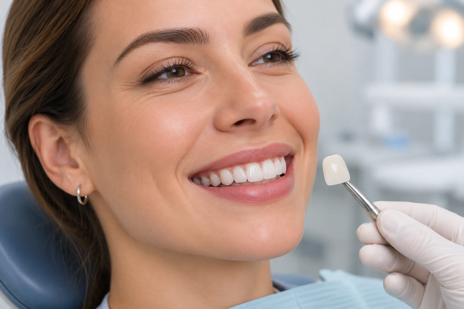
E.max veneers can chip, debond, stain at the margins, cause sensitivity, irritate the gums, or wear prematurely. Most of these problems trace back to preparation, bonding, bite alignment, or patient habits. Conservative tooth preparation, correct adhesive protocols, bite assessment, and consistent maintenance prevent the large majority of complications.
E.max veneers are thin restorations made from lithium disilicate glass ceramic. Dentists value them for their flexural strength of roughly 360 to 400 MPa, their high translucency, and their natural appearance (Rojas-Ruedas et al. 2026). Despite these strengths, E.max veneers are not indestructible. Complications arise from the clinical procedure, from the adhesive interface, and from what the patient does with the restorations every day. This article examines the most common problems with E.max veneers, explains why they occur, and gives you concrete strategies to avoid them. Each section distinguishes between a material limitation, a technical error, and a behavioural risk, because the right prevention depends on the true cause.
What Makes E.max Veneers Different From Other Porcelain Veneers?
E.max veneers contain lithium disilicate, a glass ceramic with needle-shaped crystals that resist crack propagation. This gives them greater strength than feldspathic porcelain while keeping enamel-like translucency. A veneer covers only the front surface of a tooth, unlike a crown, which covers the whole tooth.
Lithium disilicate differs from traditional feldspathic porcelain at the microstructural level. Its crystal content deflects and slows cracks, which explains its higher flexural strength (Rojas-Ruedas et al. 2026). Laboratory and clinical data show that non-feldspathic veneers, including lithium disilicate, fail less often than feldspathic veneers over long follow-up periods (Alenezi et al. 2021). Dentists therefore favour E.max for anterior teeth and selected premolars, where aesthetics and strength matter equally.
Material strength does not, however, guarantee immunity from damage. A strong ceramic still fractures when forces exceed its tolerance, and it still debonds when the adhesive interface fails. Understanding the difference between a veneer and a crown matters here as well.
Feature | E.max veneer | E.max crown |
Coverage | Front surface, sometimes wrapping the incisal edge | Entire clinical crown of the tooth |
Tooth reduction | Minimal, usually within enamel | Greater reduction on all surfaces |
Main indication | Aesthetic correction of anterior teeth | Heavily damaged or root-treated teeth |
Typical risk | Debonding, chipping at thin margins | Bulk fracture under heavy load |
What Are the Most Common Problems With E.max Veneers?
Clinical research identifies ceramic fracture and debonding as the two leading complications of porcelain veneers, followed by secondary caries and endodontic problems (Alenezi et al. 2021). Chipping, sensitivity, gum irritation, marginal staining, bite discomfort, and aesthetic dissatisfaction complete the list.
A systematic review of 25 clinical studies covering 6,500 porcelain laminate veneers found a 10-year cumulative survival rate of 95.5%. Fracture was the most common complication, and debonding followed (Alenezi et al. 2021). Each problem below has a distinct cause, a distinct prevention strategy, and a distinct treatment path.
Chipping and Cracking Under Excessive Pressure
Excessive biting force, trauma, and teeth grinding cause chips and cracks in E.max veneers. You prevent them by avoiding hard objects, protecting your bite, and wearing a night guard if you grind your teeth.
Ceramic materials resist compression well but tolerate bending and impact poorly. Biting ice, cracking nutshells, chewing hard sweets, or using your teeth to open packaging concentrates force on a small area of the ceramic edge. Accidental trauma, such as a blow to the mouth, produces the same effect. Bruxism multiplies the risk because it applies repetitive mechanical stress hundreds of times per night; clinical data show significantly higher veneer failure rates in patients who grind their teeth (Beier et al. 2012).
A dentist evaluates a minor chip differently from a full fracture. Small edge chips often respond to smoothing and polishing, while cracks that cross the restoration usually require replacement. If you notice any new edge defect, arrange an examination before the damage extends.
Veneers Becoming Loose or Falling Off
Veneer bonding failure usually results from moisture contamination during cementation, inadequate ceramic surface conditioning, or bonding to dentin instead of enamel. A loose veneer needs prompt professional assessment, not home repair.
Lithium disilicate bonds to enamel through a two-step mechanism: hydrofluoric acid etching creates microscopic retention, and a silane coupling agent creates chemical adhesion (Rojas-Ruedas et al. 2026). Any break in this chain weakens the bond. Saliva or blood contamination during cementation, incorrect etching time, or a preparation that extends into dentin all reduce adhesion, because resin cement bonds to enamel far more reliably than to dentin (Rojas-Ruedas et al. 2026). Heavy occlusal forces and trauma then finish the job by levering the veneer off the tooth.
Tooth Sensitivity After E.max Veneer Placement
Mild sensitivity to hot, cold, or sweet stimuli is common for the first days after veneer placement and usually resolves on its own. Sensitivity that persists or worsens signals a deeper problem and needs dental evaluation.
Tooth preparation removes a thin layer of enamel, which temporarily exposes the dentinal tubules to thermal and osmotic stimuli. This explains the expected short-term sensitivity. Persistent symptoms point to different causes: excessive tooth reduction that left little enamel, exposed dentin that lacks a stable bond, or pulpal irritation from deep preparation. A systematic review of porcelain veneer outcomes found that the need for root canal treatment after veneer placement remains rare, with a 10-year survival rate of 99.0% when this complication counts as failure (Alenezi et al. 2021). Conservative preparation within enamel keeps that risk low.
Gum Irritation and Inflammation Around Veneers
Poorly adapted veneer margins, residual cement, and plaque accumulation irritate the gum tissue. Redness, swelling, bleeding, and tenderness around a veneer margin warrant a dental examination.
The junction between the veneer and the tooth, called the margin, must fit precisely. An overhanging margin traps plaque that you cannot remove with a brush. Excess resin cement left below the gum line acts as a chronic irritant and promotes bacterial colonisation. Over time this produces localised gingival inflammation. Accurate margin placement, careful finishing, complete removal of cement, daily flossing, and regular professional cleaning keep the surrounding tissue healthy.
Discolouration Around Veneer Edges
The E.max ceramic itself resists staining, but the bonding margin and the natural tooth structure can discolour over time. Good oral hygiene and routine examinations limit this risk.
Lithium disilicate is a dense, glazed ceramic with very low porosity, so coffee, tea, and tobacco affect it far less than they affect natural enamel. The vulnerable zone is the margin, where the resin cement meets the tooth. Plaque, smoking, and chromogenic foods stain this interface and produce a visible dark line near the gum. Marginal staining differs from intrinsic ceramic discolouration, which is uncommon. Severe marginal discoloration that cannot be polished away is one of the recognised reasons for veneer replacement in long-term studies (Gresnigt et al. 2019).
Bite Misalignment and Discomfort When Chewing
Incorrect veneer thickness or positioning creates uneven biting forces, which cause chewing discomfort, premature contact, and jaw fatigue. Careful occlusal adjustment and digital planning prevent this problem.
When a veneer sits slightly too high, it receives the first contact every time you close your mouth. This premature contact concentrates force on one restoration instead of distributing it across the dental arch. Symptoms include discomfort when chewing, a feeling that the tooth touches first, and muscle fatigue in the jaw. Sustained uneven loading also raises the risk of ceramic fracture, because it subjects one veneer to forces designed for many teeth. Dentists prevent this through bite assessment, articulating paper checks, and progressive occlusal adjustment before final cementation.
Unnatural Appearance, Incorrect Colour, or Shape
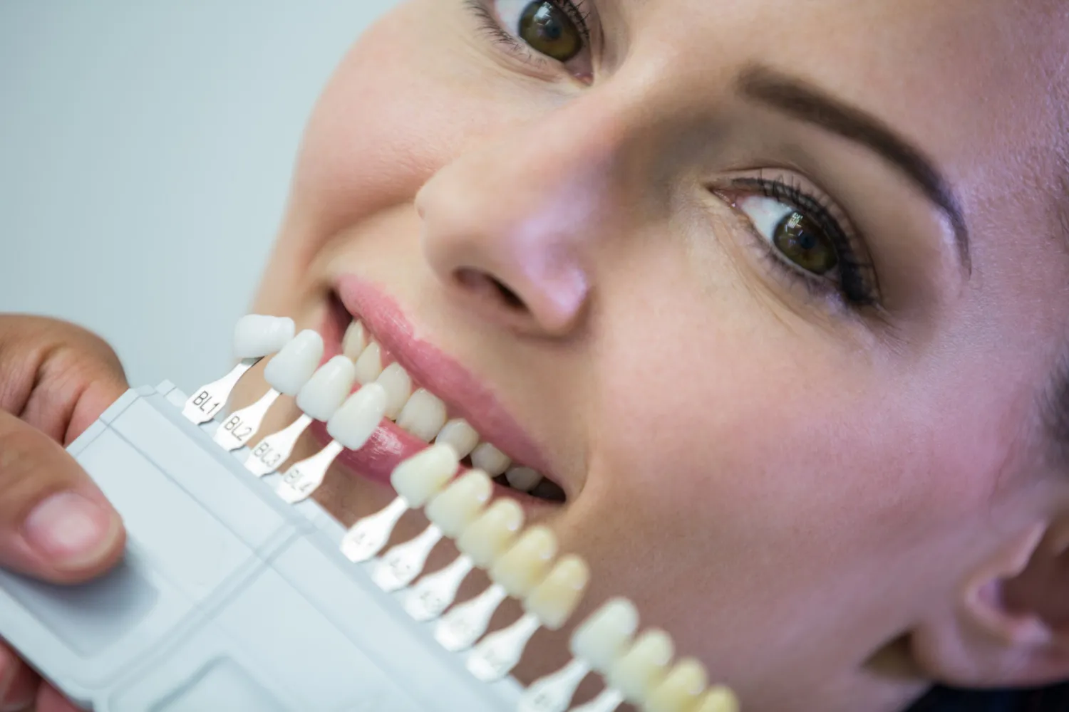
Wrong shade selection, excessive brightness, poor translucency, or incorrect proportions make veneers look artificial. A trial mock-up and your approval before bonding prevent aesthetic disappointment.
Aesthetic failure differs from mechanical failure, but it frustrates patients just as much. A shade that is too bright, a surface that is too opaque, or teeth that ignore the proportions of the face produce an unnatural smile. Shade selection must account for the neighbouring teeth, the underlying tooth colour, and the translucency of the chosen ceramic. Poor communication between patient, dentist, and laboratory causes most of these mismatches, which is why mock-ups and digital smile previews play a central role in modern planning (Rojas-Ruedas et al. 2026).
Tooth Decay Developing Beneath E.max Veneers
Ceramic cannot decay, but the natural tooth underneath remains vulnerable to cavities. Plaque at the veneer margin and poor flossing habits drive this risk.
Secondary caries forms when bacteria colonise the junction between veneer and tooth, especially where the margin fits poorly or where cement has washed out. Marginal leakage allows fluids and bacteria to reach the underlying dentin. The good news is that this complication is rare: a large systematic review reported a 10-year survival rate of 99.3% when secondary caries alone counts as failure, with most cases appearing only after five years (Alenezi et al. 2021). Daily brushing, daily flossing, and regular margin checks keep this risk minimal.
Premature Wear and Loss of Surface Gloss
Abrasive toothpaste, aggressive brushing, and contact with opposing teeth gradually dull the ceramic surface. A soft-bristled brush and a non-abrasive toothpaste preserve the glaze.
The glaze on a ceramic veneer gives it a lifelike lustre and a smooth surface that resists plaque. Abrasive oral care products scratch this layer over time, and constant contact with opposing natural teeth adds further wear. A dull veneer collects more plaque, which connects surface wear back to the gum inflammation and marginal staining described earlier. If professional polishing becomes necessary, the dentist should use ceramic-specific polishing systems rather than standard prophylaxis pastes.
Why Do E.max Veneer Complications Occur?
Complications arise from four main sources: inadequate tooth preparation, incorrect adhesive technique, poor case selection, and insufficient bite assessment. Identifying the true cause matters more than treating only the visible damage.
Every complication described above has an origin in one of these four categories. A veneer that falls off twice is not unlucky; it usually signals a bonding or occlusal problem that the first repair did not address. Understanding the cause guides both prevention and treatment.
Inadequate Tooth Preparation and Enamel Removal
Preserving enamel is the single most important preparation principle, because the resin bond to enamel is far stronger than the bond to dentin.
Excessive reduction removes the very substrate that makes adhesive dentistry work. A preparation that reaches deep dentin produces weaker adhesion, more post-operative sensitivity, and poorer retention (Rojas-Ruedas et al. 2026). Inappropriate preparation also distorts veneer thickness, which compromises both fit and aesthetics. The clinical rule is simple: remove only what the restoration needs, and no more.
Incorrect Adhesive Selection and Cementation Techniques
Lithium disilicate requires hydrofluoric acid etching followed by silane application. Contamination or incorrect timing at any step weakens the final bond.
The etchable glass phase of lithium disilicate allows reliable micromechanical retention, but only when the laboratory and the dentist follow the correct protocol (Rojas-Ruedas et al. 2026). After etching, even brief contact with saliva contaminates the ceramic surface and reduces bond strength. Cement selection matters as well: light-cured resin cements suit thin, translucent veneers, while complete removal of excess cement protects the gum tissue discussed earlier. Proper isolation, controlled curing, and meticulous finishing complete the procedure.
Poor Case Selection and Incorrect Treatment Planning
E.max veneers do not suit every patient. Severe tooth damage, extensive existing restorations, insufficient enamel, active gum disease, and uncontrolled bruxism all require a different plan.
Case selection determines long-term success more than any brand of ceramic. Teeth with large existing fillings offer little enamel for bonding. Patients with untreated periodontal disease provide an unstable biological foundation. Heavy grinders place repetitive loads on thin ceramic edges, and bruxism correlates with higher veneer failure rates in clinical follow-up studies (Granell-Ruíz et al. 2014). In such situations, full crowns, orthodontics, or preliminary bite therapy often deliver more predictable outcomes than veneers alone.
Insufficient Bite Assessment Before Placement
An untreated bite discrepancy concentrates pressure on individual veneers and accelerates failure. Occlusion, jaw movement, and the opposing teeth all belong in the treatment plan.
A veneer does not exist in isolation. It functions within a system of contacts, guidance movements, and muscle forces. If the plan ignores this system, even a perfectly bonded veneer will carry loads it was never designed for. Digital treatment planning and a thorough occlusal analysis allow the dentist to balance aesthetic goals with functional requirements before any tooth is prepared.
How Can You Prevent Problems Before Getting E.max Veneers?

Prevention starts before treatment. Choose an experienced dentist, resolve existing oral health problems, approve a previewed smile design, and insist on conservative preparation.
The four failure sources above translate directly into four preventive actions. Each one addresses a cause before it can produce a complication.
Choose a Dentist Experienced in Lithium Disilicate Restorations
The operator's skill influences veneer survival as much as the material. Verify experience with E.max cases before you commit.
Ask how many lithium disilicate veneer cases the dentist has completed, and review genuine before-and-after photographs. Ask which dental laboratory fabricates the restorations and which ceramic system the laboratory uses. A competent clinician will also discuss limitations, possible complications, and maintenance openly rather than presenting veneers as a risk-free procedure.
Evaluate Your Oral Health Before Treatment
Treat cavities, gum disease, and infections first. Veneers placed on an unhealthy foundation fail early.
A pre-treatment assessment should cover existing decay, periodontal status, tooth sensitivity, previous restorations, and grinding habits. This evaluation answers a more basic question as well: whether veneers are the right treatment at all. Sometimes whitening, orthodontics, or composite bonding solves the aesthetic problem with less intervention.
Approve the Smile Design Before Permanent Bonding
Preview the planned shape, shade, and proportions with a mock-up or digital design, and confirm your approval before any irreversible step.
Digital smile design and temporary mock-ups let you test the proposed result in your own mouth. You can evaluate tooth length, shape, and colour against your facial features and your remaining natural teeth. This step converts aesthetic expectations into an agreed plan, which prevents the dissatisfaction described in the section on unnatural appearance.
Prioritise Conservative Tooth Preparation
Every micrometre of preserved enamel improves bonding, reduces sensitivity, and supports a natural look.
This point closes the loop with the causes section: because enamel preservation drives bond strength, conservative preparation is not a compromise but a clinical advantage. Long-term data support this approach, with 10-year survival rates above 96% reported for lithium disilicate veneers placed with enamel-focused protocols (Klein et al. 2025).
How Should You Care for E.max Veneers to Extend Their Lifespan?
Brush twice daily with a soft brush, floss daily, avoid hard objects and parafunctional habits, protect against grinding, and attend regular examinations.
Maintenance converts a good clinical result into a durable one. The routine below targets each complication category without adding unnecessary steps.
Follow a Daily Oral Hygiene Routine
Two minutes of brushing twice a day with a soft-bristled brush and non-abrasive toothpaste, plus daily flossing, protects both the veneer margins and the tooth underneath.
Flossing cleans the interproximal areas where veneer margins meet neighbouring teeth, which are the zones where plaque triggers gum inflammation and secondary caries. Healthy gums, in turn, stabilise the tissue frame that keeps the margins looking natural.
Avoid Foods and Habits That Can Damage Ceramic Veneers
Do not bite ice, hard sweets, or nutshells, and never use your teeth as tools. These habits generate the concentrated forces that chip ceramic edges.
Sticky foods deserve caution as well, because they pull on the restoration while you chew. Nail biting and pen chewing create the same repetitive micro-trauma that grinding produces, only during waking hours.
Protect Veneers From Nighttime Teeth Grinding
A custom-made night guard absorbs grinding forces and is the standard protective measure for patients with bruxism.
Bruxism applies repetitive mechanical stress that accumulates as fatigue damage in the ceramic. Research on porcelain veneers confirms that patients with bruxism face a significantly higher failure rate, and that occlusal splints modify this risk (Beier et al. 2012). Jaw pain, morning headaches, or visible tooth wear are warning signs that justify a professional assessment and, where indicated, a night guard.
Attend Regular Dental Examinations
Routine reviews detect marginal defects, early chips, and bite changes before they become failures.
At each visit the dentist examines the veneer margins, the bonding integrity, the gum health, and the occlusion. Professional cleaning removes deposits that home care misses. These examinations also protect the natural teeth beneath the restorations, which remain biological structures with their own needs.
What Should You Do If Your E.max Veneers Develop Problems?
Contact your dentist promptly, keep any detached veneer in a safe container, and never attempt repairs or reattachment yourself. The right treatment depends on a professional diagnosis.
Most veneer problems have a workable solution when you act early. The sections below cover the four situations patients ask about most.
Can a Chipped E.max Veneer Be Repaired?
Minor chips can sometimes be polished or repaired with composite, but cracks that compromise the ceramic usually require replacement.
A small edge chip that does not affect aesthetics or function may only need smoothing. Deeper damage, or any visible crack, makes replacement the more predictable option because a repaired ceramic edge remains a weak point. Never file or polish a veneer yourself: household abrasives destroy the glaze and create a rough surface that traps plaque.
What Happens If an E.max Veneer Falls Off?
Store the detached veneer in a clean container and see your dentist quickly. Do not use household adhesives under any circumstances.
A detached veneer that is intact can sometimes be re-bonded, but only after the dentist cleans both surfaces and checks the underlying tooth and the original bonding protocol. Household glues are toxic, unpredictable, and contaminate the ceramic, which usually destroys any chance of professional reattachment. A prompt visit also protects the exposed prepared tooth from sensitivity and decay.
How Are Persistent Sensitivity and Pain Treated?
Persistent symptoms require a clinical examination and, where needed, diagnostic tests. Treatment follows the diagnosis, not the veneer material.
Possible causes include exposed dentin, a bite discrepancy, pulp inflammation, or decay beneath the restoration. Each cause has a different treatment, from a simple occlusal adjustment to endodontic therapy. This is why self-diagnosis fails: the symptom tells you that something is wrong, but only an examination tells you what.
When Do E.max Veneers Need Replacement?
Visible cracks, extensive fractures, repeated debonding, poor marginal fit, and unacceptable aesthetic deterioration justify replacement. There is no fixed replacement schedule.
The decision weighs the condition of the ceramic against the condition of the tooth beneath it. A veneer with a failing margin on a healthy tooth is a straightforward replacement. A veneer over a decayed or fractured tooth needs broader treatment. Long-term studies show that veneers individually fail at different times, so replacement timing varies from patient to patient rather than following a calendar (Fradeani et al. 2005).
How Long Do E.max Veneers Last Without Complications?
Published clinical studies report survival rates above 95% at ten years for lithium disilicate veneers, provided case selection, bonding, and maintenance are correct. These figures describe groups, not individual guarantees.
A systematic review and meta-analysis reported a 96.81% survival rate for lithium disilicate veneers over a mean follow-up of 10.4 years, with fewer technical and aesthetic complications than feldspathic or leucite-reinforced ceramics (Klein et al. 2025). A separate 10-year clinical study recorded a cumulative survival rate of 96.3% for lithium disilicate veneers in anterior aesthetic cases (Fradeani et al. 2005). Real-world dental veneer longevity depends on oral hygiene, bite forces, bonding quality, diet, and regular professional care. It also helps to separate two concepts: the ceramic material itself is chemically stable for decades, while the bonded restoration as a system ages at its interfaces. Maintenance targets those interfaces.
Are E.max Veneers Worth It Despite Their Potential Problems?
For the right patient, yes. E.max veneers combine natural translucency, stain resistance, and strong long-term survival data, but they demand irreversible preparation, careful case selection, and ongoing maintenance.
Aspect | E.max (lithium disilicate) | Feldspathic porcelain | Composite veneers |
Strength | High, 360 to 400 MPa | Lower, more brittle | Lowest |
Aesthetics | High translucency, layered options | Excellent, very lifelike | Good, degrades over time |
Stain resistance | Very high | High | Moderate |
Repairability | Limited, often replacement | Limited | Easy chairside repair |
Long-term survival | Above 95% at 10 years | Lower than non-feldspathic ceramics | Shorter lifespan |
The limitations are real. Treatment costs more than composite alternatives, preparation removes enamel permanently, and unsuitable cases fail early. A systematic review comparing feldspathic porcelain with glass-ceramic veneers found that glass ceramics offer the better complication profile, which supports E.max for patients who meet the clinical criteria (Morimoto et al. 2016). An individual assessment matters more than any material comparison, because the best ceramic in the wrong mouth still fails.
Frequently Asked Questions About E.max Veneer Problems
The questions below summarise the concerns patients raise most often. Each answer links back to the detailed sections above.
Can E.max Veneers Crack Like Natural Teeth?
Yes. Ceramic fractures under excessive pressure or trauma, just as enamel does.
The difference lies in the threshold and in protection. Sound case selection, a balanced bite, and a night guard for grinders reduce fracture risk to the low single digits observed in long-term studies (Alenezi et al. 2021).
Do E.max Veneers Cause Bad Breath?
The ceramic itself does not cause bad breath, but plaque at poorly adapted margins and inflamed gums can.
Food debris and bacterial biofilm around a defective margin produce the odour. Correct margin fit, daily flossing, and professional cleaning eliminate the source rather than masking it.
Can E.max Veneers Damage Natural Teeth?
Properly planned veneers preserve tooth structure. Damage arises from excessive preparation, poor bonding, untreated decay, or a misaligned bite.
The procedure aims to stay within enamel precisely because this protects the tooth. When a complication does reach the natural tooth, the cause usually sits in the planning or maintenance chain rather than in the ceramic.
Do E.max Veneers Turn Yellow Over Time?
No. Lithium disilicate is highly stain resistant and keeps its intrinsic colour.
What patients perceive as yellowing is usually staining at the margins or darkening of the natural tooth structure beside the veneer. This is a hygiene and margin-quality issue, not a material defect.
Can You Whiten E.max Veneers After They Become Discoloured?
No. Whitening gels act on natural tooth structure and cannot change the intrinsic shade of ceramic.
Professional cleaning removes surface deposits and restores the original appearance in most cases. When the underlying tooth has darkened and shows through the translucent ceramic, replacement with a new shade is the predictable solution.
Is It Normal to Experience Sensitivity After Getting E.max Veneers?
Mild, short-lived sensitivity after preparation and bonding is normal. Sensitivity that persists beyond the first weeks or worsens needs assessment.
The temporary response reflects enamel removal and dentinal exposure during preparation. Ongoing symptoms point to the causes discussed in the sensitivity section and justify a clinical review.
Can You Eat Normally With E.max Veneers?
Yes, after the initial recovery period and your dentist's instructions. Normal eating is safe; extreme biting forces are not.
The practical rule is to treat veneers as you should treat natural teeth: avoid biting ice, hard sweets, and non-food objects, and let the night guard handle the forces you cannot control.
Final Perspective on Common Problems With E.max Veneers and How to Avoid Them
The common problems with E.max veneers follow predictable patterns. Fracture and debonding lead the list, marginal and biological complications follow, and nearly all of them trace back to preparation, bonding, case selection, bite management, or daily habits. The evidence is reassuring: with conservative preparation, correct adhesive protocols, and structured maintenance, lithium disilicate veneers achieve survival rates above 95% at ten years (Klein et al. 2025). Your role is to choose an experienced clinician, protect the restorations from avoidable forces, and keep every review appointment. That combination addresses each complication at its true source.
References
Alenezi, A., et al. "Long-Term Survival and Complication Rates of Porcelain Laminate Veneers in Clinical Studies: A Systematic Review." Journal of Clinical Medicine, vol. 10, no. 5, 2021, article 1074.
Beier, U. S., et al. "Clinical Performance of Porcelain Laminate Veneers for up to 20 Years." The International Journal of Prosthodontics, vol. 25, no. 1, 2012, pp. 79-85.
Fradeani, M., et al. "Porcelain Laminate Veneers: 6- to 12-Year Clinical Evaluation. A Retrospective Study." The International Journal of Periodontics and Restorative Dentistry, vol. 25, no. 1, 2005, pp. 9-17.
Granell-Ruíz, M., et al. "Influence of Bruxism on Survival of Porcelain Laminate Veneers." Medicina Oral, Patología Oral y Cirugía Bucal, vol. 19, no. 4, 2014, pp. e426-e432.
Klein, P., et al. "Survival and Complication Rates of Feldspathic, Leucite-Reinforced, Lithium Disilicate and Zirconia Ceramic Laminate Veneers: A Systematic Review and Meta-Analysis." Journal of Esthetic and Restorative Dentistry, vol. 37, no. 3, 2025, pp. 601-619.
Morimoto, S., et al. "Main Clinical Outcomes of Feldspathic Porcelain and Glass-Ceramic Laminate Veneers: A Systematic Review and Meta-Analysis of Survival and Complication Rates." The International Journal of Prosthodontics, vol. 29, no. 1, 2016, pp. 38-49.
Rojas-Ruedas, S., et al. "Narrative Review of Lithium Disilicate Veneers in Esthetic Dentistry: Current Perspectives and Clinical Case Report." Dentistry Journal, 2026.