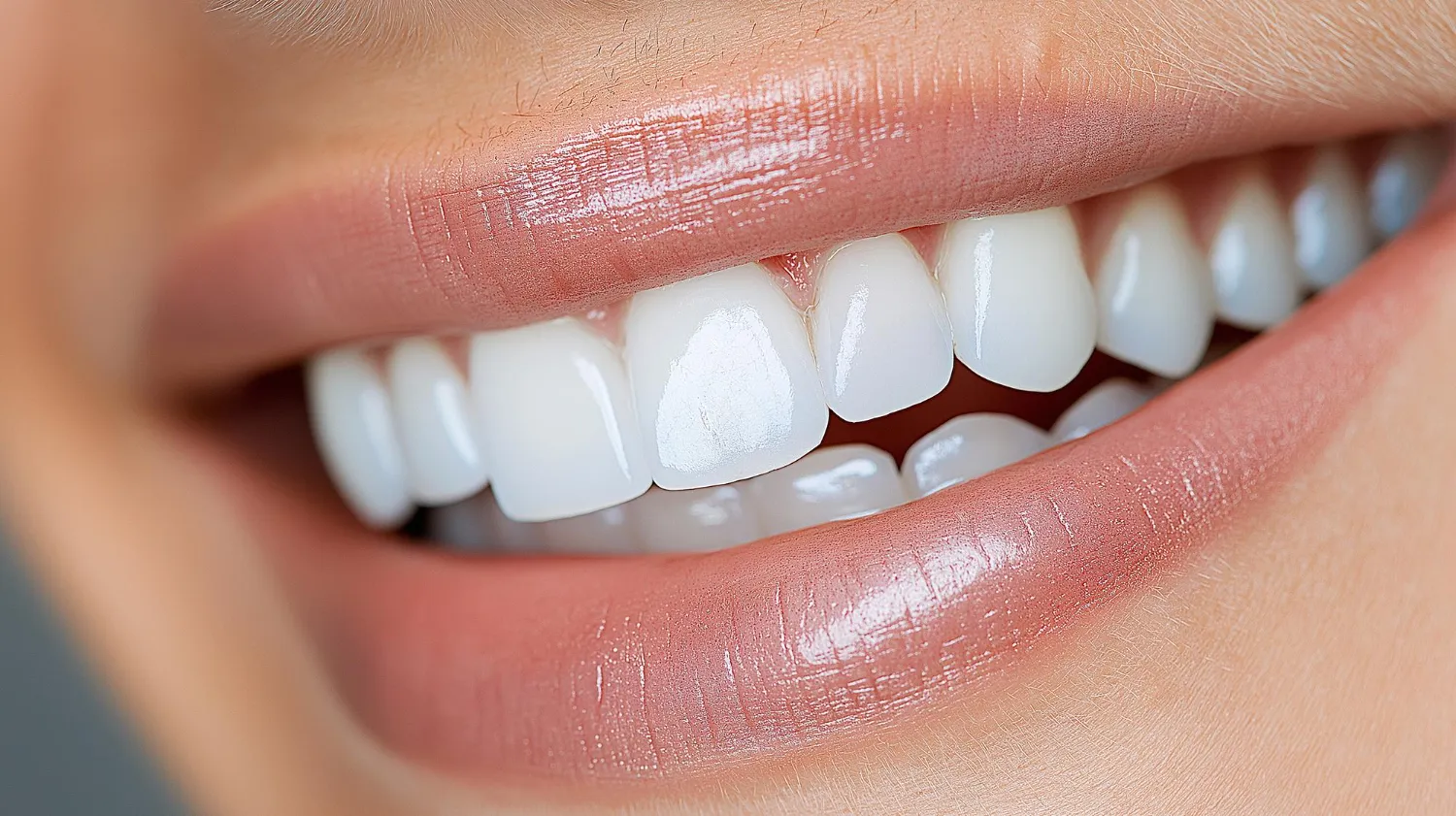
E.max veneers last 10 to 15 years on average. With excellent care, they can last 20 years or more. Clinical studies report survival rates above 90% at the 10-year mark. Your daily habits, your dentist's skill, and the quality of the laboratory work all shape how long your veneers stay in place. This article breaks down every factor that affects E.max veneer longevity. We draw from peer-reviewed clinical studies to give you real answers.
What Are E.max Veneers?
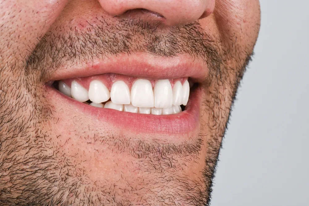
E.max veneers are thin ceramic shells. Dentists bond them to the front surfaces of teeth. They improve color, shape, and alignment. E.max is a brand name. It refers to IPS E.max, a lithium disilicate glass-ceramic material made by Ivoclar Vivadent. Dentists use E.max for veneers, crowns, inlays, and onlays. The material has gained massive popularity in cosmetic dentistry over the past two decades.
What Is IPS E.max Lithium Disilicate?
IPS E.max lithium disilicate is a glass-ceramic material. It contains lithium disilicate crystals embedded in a glassy matrix. This composition gives the material both strength and beauty. Al-Thobity and Alsalman (2021) tested three lithium disilicate materials in their study. They found that IPS E.max CAD reached a flexural strength of 364.64 MPa. IPS E.max Press reached 249.59 MPa. Both values exceed the ISO 6872 requirements for clinical use. Zulkiffli et al. (2024) confirmed similar results. They reported a flexural strength of 381.12 MPa for IPS E.max CAD. These numbers place E.max well above older ceramic materials like feldspathic porcelain.
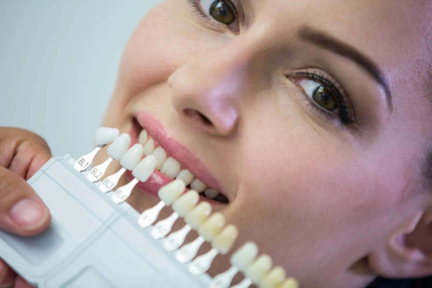
The optical properties of E.max also stand out. The material comes in multiple translucency levels. These include high translucency (HT), medium translucency (MT), low translucency (LT), and medium opacity (MO). Willard (2018) explains that lithium disilicate achieves its translucency through a matching refractive index between the crystalline and glassy phases. This property allows light to pass through the veneer much like it passes through natural enamel. The result is a restoration that looks alive, not flat or artificial.
Why Do Dentists and Patients Prefer E.max?
Dentists prefer E.max for several reasons. First, the material offers natural translucency. Patients want veneers that look like real teeth, not like white blocks. E.max delivers this esthetic quality. Second, E.max allows conservative tooth preparation. Dentists can place E.max veneers with minimal enamel removal. Some preparations require only 0.3 mm of reduction. This conservatism preserves healthy tooth structure. Third, E.max shows excellent biocompatibility. Research by Malchiodi et al. (2019) evaluated 79 lithium disilicate multilayered anterior veneers over three years. They reported a 98.7% survival rate and noted high patient satisfaction with both esthetics and comfort. Fourth, the material bonds reliably to enamel. This strong adhesive connection contributes directly to long-term success.
How Long Do E.max Veneers Last?
E.max veneers typically last 10 to 15 years. Many patients keep their veneers for 20 years or longer. Clinical data supports these claims with hard numbers. The exact lifespan depends on the patient, the dentist, and the care routine.
What Is the Average Lifespan?
Most dental professionals estimate a 10-to-15-year lifespan for E.max veneers. This estimate aligns with clinical evidence. Demirekin et al. (2022) conducted a retrospective study on IPS E.max Press veneers. They reported a remarkable 99.7% survival rate after 10 years. Aslan et al. (2019) analyzed lithium disilicate laminate veneers placed by experienced dentists. They found a 97.4% survival rate at the 10-year follow-up. These numbers suggest that the vast majority of E.max veneers remain intact and functional for at least a decade.
Some patients enjoy even longer service life. Layton and Walton (2007) conducted a prospective study of porcelain veneers with up to 16 years of follow-up. Their data showed Kaplan-Meier cumulative survival rates of 96% at 10 years and 91% at 15 years. Fradeani et al. (2005) performed a retrospective evaluation of porcelain laminate veneers over 6 to 12 years. Their results confirmed high success rates with proper case selection and technique. While these studies included various ceramic types, they establish a clear pattern. Well-placed ceramic veneers, including E.max, can last well beyond 15 years.
What Do Clinical Survival Rates Show?
Clinical survival rates give us the most reliable picture of veneer longevity. Survival rate means the percentage of veneers still in place and functioning at a given time point. Morimoto et al. (2016) performed a systematic review and meta-analysis of feldspathic porcelain and glass-ceramic laminate veneers. They reported an estimated overall cumulative survival rate of 89% at a median follow-up of 9 years. For glass-ceramic veneers specifically, which include E.max, the survival rate reached 94%.
Klein et al. (2021) conducted another systematic review and meta-analysis. They analyzed survival and complication rates of various ceramic veneer materials. Their results showed a 96.8% survival rate at 10.4 years for ceramic laminate veneers. They specifically noted that lithium disilicate veneers may be preferred as a restorative material for long-term success. Imburgia et al. (2021) tracked 1,075 lithium disilicate CAD/CAM veneers placed on 105 patients. They reported an outstanding 99.83% survival rate over a mean observation period of 30.8 months. Nejatidanesh et al. (2018) followed chairside CAD/CAM lithium disilicate veneers for five years. They recorded a 97.8% survival rate.
Fotiadou et al. (2021) evaluated lithium disilicate indirect restorations over 8.5 years. They calculated a 94% survival rate at that extended time point. Rinke et al. (2020) reported a 10-year survival rate of 91.8% for ceramic veneers in their clinical practice. These studies span different countries, operators, and patient populations. Yet they all point to the same conclusion. E.max veneers demonstrate excellent long-term survival when clinicians apply proper technique and patients maintain good oral health.
Can E.max Veneers Last a Lifetime?
E.max veneers can last a lifetime, but this outcome is not guaranteed. No clinical study has tracked E.max veneers for a full human lifetime. The longest published follow-ups reach 14 to 16 years. AlJazairy (2020) conducted a systematic review of porcelain laminate veneer survival. He noted that conclusive estimation of longevity beyond 20 years remains lacking. The dropout rate in long-term studies becomes exceedingly high after two decades. This gap in data makes lifetime predictions speculative.
However, individual cases suggest that decades of service are possible. Olley et al. (2018), cited in AlJazairy's review, reported on ceramic veneers in a dental practice with up to 50 years of follow-up. They recorded 100% survival for 22 feldspathic porcelain veneers at the 50-year mark. While this study involved a small sample and older materials, it proves that ceramic veneers can endure for half a century under ideal conditions. For E.max specifically, the material's superior flexural strength and fracture toughness compared to traditional feldspathic porcelain suggest that comparable or better longevity is achievable.
Realistic expectations matter. Patients should view E.max veneers as a long-term investment, not a permanent solution. With excellent care, regular dental visits, and protective habits, many patients will enjoy their E.max veneers for 20 years or more. Some may never need replacement. Others might face earlier failure due to trauma, poor bonding, or untreated bruxism.
What Determines the Lifespan of E.max Veneers?
Five main factors determine how long E.max veneers last. These include the dentist's experience, the bonding technique, the dental laboratory quality, the patient's oral health, and the bite forces acting on the veneers. Each factor plays a distinct role. Together, they create the conditions for either success or early failure.
How Does the Dentist's Skill Affect Longevity?
The dentist's skill shapes every aspect of veneer treatment. First, the dentist must perform proper case selection. Not every patient qualifies for veneers. Teeth with large existing fillings, severe decay, or poor periodontal health may need alternative treatments. Second, the dentist must design the smile correctly. This design includes tooth proportions, incisal edge position, and contact points. Poor design leads to functional problems and esthetic dissatisfaction. Third, tooth preparation requires precision. The dentist must remove enough enamel to allow proper veneer thickness. At the same time, the dentist must preserve as much healthy tooth structure as possible. Over-preparation weakens the tooth. Under-preparation creates a bulky veneer that feels unnatural and traps plaque.
Gonzalez-Martin et al. (2021) investigated factors affecting veneer fracture. They found that preparation type significantly influenced outcomes. Their generalized estimating equation model revealed that the odds of veneer fracture were significantly higher in patients who exhibited tooth wear at baseline. This finding underscores the importance of thorough pre-treatment assessment. An experienced dentist identifies these risk factors before placing the first veneer.
Why Does Bonding Technique Matter?
Bonding technique directly determines whether a veneer stays attached or falls off. Adhesive cementation creates a micromechanical and chemical bond between the ceramic, the resin cement, and the tooth structure. This process demands strict protocol. The dentist must etch the enamel with phosphoric acid. Then the dentist applies a bonding agent. The dentist must also etch the internal surface of the E.max veneer with hydrofluoric acid and apply silane coupling agent. This step creates a chemical bridge between the glass-ceramic and the resin cement.
Proper isolation prevents contamination. Saliva, blood, or gingival fluid on the bonding surface ruins the adhesive connection. Even a tiny amount of moisture can cause debonding within months. The dentist must maintain a dry field throughout the cementation procedure. Long-term bond strength depends on every detail of this process. A 2024 meta-analysis published in the Journal of Prosthetic Dentistry examined ceramic veneers bonded to different substrates. The study found that enamel-bonded veneers achieved almost perfect survival rates of 99%. Veneers bonded to exposed dentin showed significantly lower survival rates of 91%. This dramatic difference highlights why conservative enamel preparation and meticulous bonding technique matter so much.
How Does Lab Quality Influence Durability?
The dental laboratory fabricates the actual veneer. Lab quality affects fit, thickness, and internal integrity. Modern labs use CAD/CAM technology to mill E.max veneers from pre-crystallized blocks. After milling, the technician heat-treats the veneer in a furnace. This crystallization process develops the final strength, shade, and translucency. Precision matters at every stage. A poorly milled veneer may have internal flaws. These flaws act as stress concentrators. Under chewing forces, cracks propagate from these flaws and cause fracture.
Sen et al. (2024) evaluated CAD/CAM laminate veneers fabricated from either lithium disilicate (E.max CAD) or feldspathic ceramic (VMII). They followed these veneers for up to 10 years. The E.max CAD veneers showed a 92.7% survival rate. The feldspathic veneers showed an 89.1% survival rate. While both materials performed well, the lithium disilicate group had a slight edge. The researchers attributed this difference to the material's superior mechanical properties. A high-quality lab maximizes these material advantages through precise fabrication.
What Role Does Oral Health Play?
The patient's oral health creates the biological environment for the veneers. Healthy gums form a tight seal around the veneer margins. This seal protects the underlying tooth from bacterial invasion. Inflamed or receding gums expose the margin. Bacteria accumulate in this gap. Over time, this leads to secondary decay or periodontal disease. Research published in PMC (2024) evaluated E.max porcelain veneers over five years. The study examined 28 patients with 6-to-10-unit veneer cases. They found that 64.3% of patients displayed healthy gingival status with no bleeding. However, 35.7% showed inflamed gingival tissue. The majority of these patients had no significant veneer changes like marginal staining, debonding, or chipping. Still, the presence of inflammation signals a risk factor for long-term problems.
Existing restorations on the tooth also affect outcomes. A tooth with a large composite filling provides a weaker bonding substrate than intact enamel. The 2024 substrate meta-analysis confirmed this. Veneers bonded to composite resin or surfaces with minimal dentin exposure showed survival rates of 94%. This rate drops further when severe dentin exposure occurs. Patients with multiple existing fillings need careful evaluation before veneer placement.
How Do Bite Forces Impact Veneer Survival?
Bite forces represent the mechanical challenge that veneers face every day. Normal chewing generates predictable loads. Parafunctional habits generate destructive forces. Bruxism, or teeth grinding, applies lateral and compressive forces that far exceed normal function. Clenching creates sustained high pressure. These habits transfer stress to the veneer edges. The incisal edge of a veneer is the thinnest and most vulnerable area. Repeated stress causes microcracks. Eventually, these cracks grow into visible chips or complete fractures.
A study published in PMC examined the influence of bruxism on porcelain laminate veneer survival. Researchers placed 323 veneers in 70 patients. They found that bruxism activity significantly increased the risk of failure. Of the 13 fractures and 29 debonding incidents observed, 8 fractures and 22 debonding cases were related to bruxism. The researchers concluded that occlusal splints reduce the risk of fractures in bruxism patients. Gonzalez-Martin et al. (2021) also found that men had significantly higher odds of veneer fracture than women. Patients with tooth wear at baseline showed 5.54 times higher odds of fracture. Central and lateral incisors fractured more often than canines and premolars. These findings confirm that bite forces and parafunctional habits play a major role in veneer longevity.
What Daily Habits Extend the Life of E.max Veneers?
Your daily habits either protect your E.max veneers or wear them down. Small choices add up over years. Patients who adopt protective habits enjoy significantly longer veneer lifespans.
What Oral Hygiene Routine Works Best?
Brush your teeth twice daily with a soft-bristled toothbrush. Use non-abrasive toothpaste. Abrasive pastes scratch the ceramic surface. These scratches create areas where stains accumulate. Floss daily to remove plaque from between teeth and along the gumline. Plaque buildup at the veneer margin causes gingival inflammation. Inflammation leads to gum recession and margin exposure. Schedule professional cleanings every six months. Your hygienist uses specialized tools that clean thoroughly without damaging the veneer surface.
Why Should Bruxism Patients Wear a Night Guard?
Patients with bruxism must wear a custom night guard. This device creates a protective barrier between the upper and lower teeth. It absorbs grinding forces. It prevents direct contact between the veneer and the opposing teeth. Faus-Matoses et al. (2020) conducted an 8-year prospective clinical investigation on feldspathic veneers. They specifically studied the influence of occlusal splints in patients with bruxism. Their findings confirmed that night guards substantially extend veneer survival. Without a night guard, bruxism patients face fracture and debonding risks that multiply over time. With a night guard, these patients can achieve survival rates comparable to non-bruxers.
Which Foods Protect Veneers?
Choose foods that do not challenge the veneer's structural integrity. Soft foods, cooked vegetables, and normal-textured meals pose no threat. Cut hard foods like apples and carrots into pieces before eating. Do not bite directly into hard items with your front teeth. Limit sticky candies and extremely hard nuts. These foods can generate sudden high forces. They can also pull at the veneer margins. Avoid excessive acidic foods and drinks. Acid does not harm the ceramic itself, but it can erode the exposed enamel at the veneer margin. This erosion compromises the seal and invites decay.
What Habits Should Patients Avoid?
Never use your teeth as tools. Do not open packages, tear tape, or crack nuts with your veneers. These actions concentrate force on small areas. They cause chips and cracks. Stop nail biting. This habit applies repeated lateral forces to the incisal edges. It also introduces bacteria to the mouth. Do not chew ice, pens, or hard candy. Each bite risks a fracture. Do not smoke. Smoking stains the resin cement and the exposed tooth structure at the margin. It also reduces blood flow to the gums. This reduction impairs healing and increases periodontal risk.
Why Do E.max Veneers Fail Early?
Early failure frustrates both patients and dentists. Understanding the causes helps patients prevent them. The most common failure modes include fracture, debonding, secondary decay, gum recession, trauma, and poor oral hygiene.
What Causes Veneer Fracture or Chipping?
Fracture is the leading cause of veneer failure. Morimoto et al. (2016) reported that fracture and chipping accounted for approximately 4% of all ceramic veneer failures over 10 years. The incisal edge is the most common fracture site. This area bears the highest functional load. It is also the thinnest part of the veneer. Bruxism, biting hard objects, and traumatic occlusion all contribute to fracture. A 2024 meta-analysis confirmed that fracture remains the primary cause of veneer failure across multiple studies. The good news is that most fractures are preventable. Night guards, dietary awareness, and proper bonding technique eliminate the majority of fracture risk.
What Is Debonding?
Debonding means the veneer comes loose or falls off completely. This failure mode ranks second after fracture. Morimoto et al. (2016) reported debonding at approximately 2% over 10 years. Most debonding incidents occur within the first two years. They almost always result from technique errors during cementation. Insufficient acid etching, missing silane application, moisture contamination, or bonding to dentin instead of enamel all cause debonding. The veneer itself rarely causes the problem. The adhesive interface does. An experienced dentist who follows strict isolation and bonding protocols can reduce debonding rates to near zero.
Can Teeth Decay Under Veneers?
Yes, teeth can decay under veneers. This condition is called secondary caries. It develops at the margin where the veneer meets the tooth. Bacteria penetrate microscopic gaps. They produce acid. The acid dissolves the enamel and dentin beneath the veneer. The patient often does not notice early decay because the veneer covers it. Regular dental checkups catch this problem before it spreads. Morimoto et al. (2016) reported secondary caries as a failure mode in approximately 1% of cases over 10 years. Good oral hygiene and professional cleanings keep this risk minimal.
How Does Gum Recession Affect Veneers?
Gum recession exposes the veneer margin. This exposure creates both an esthetic problem and a biological risk. The margin becomes visible as a line where the ceramic meets the tooth. Stains accumulate here. Bacteria colonize the exposed cement layer. Over time, this leads to inflammation, decay, or veneer failure. Recession often results from periodontal disease, aggressive brushing, or anatomical factors. Patients with thin gingival biotypes face higher recession risk. Maintaining healthy gums through gentle brushing and regular cleanings protects both the tissue and the veneer.
What Role Does Trauma Play?
Physical trauma causes sudden, catastrophic veneer failure. Sports injuries, falls, and accidents can fracture or knock off a veneer instantly. Trauma accounts for a small percentage of overall failures, but it produces the most dramatic results. Patients who play contact sports should wear mouthguards. This simple device absorbs impact forces. It protects both the veneers and the natural teeth underneath.
Does Poor Oral Hygiene Cause Failure?
Poor oral hygiene accelerates every other failure mode. Plaque buildup causes gingivitis. Gingivitis progresses to periodontitis. Periodontitis destroys the bone and gum support around the tooth. As the gum recedes, the veneer margin becomes compromised. Plaque also harbors acid-producing bacteria. These bacteria attack the tooth structure at the margin. The 5-year E.max study mentioned earlier found that patients with inflamed gingival tissue showed higher biological risk. Proper brushing, flossing, and professional maintenance prevent this cascade.
What Signs Show E.max Veneers Need Replacement?
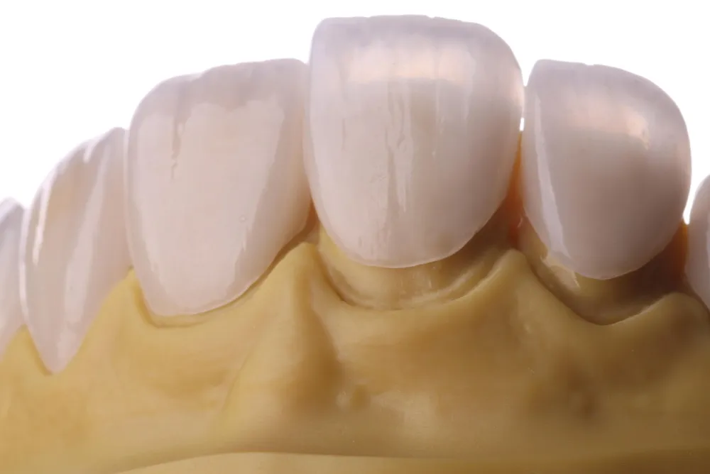
Veneers do not last forever. Patients should watch for warning signs. Early detection allows for timely replacement. Delayed action risks damage to the underlying tooth.
How Do Chips or Cracks Appear?
Visible chips or cracks are the most obvious sign of veneer failure. Small chips at the incisal edge may seem minor. However, they indicate that the material has reached its stress limit. Cracks can propagate inward. They can eventually cause complete fracture. Any visible damage warrants a dental evaluation. Your dentist can determine whether repair or replacement is appropriate.
What Is Marginal Staining?
Marginal staining appears as a brown or gray line at the edge of the veneer. It indicates that the cement layer has discolored. It may also signal microleakage. Microleakage means that fluids and bacteria are seeping under the veneer margin. This seepage leads to secondary decay. Marginal staining is both an esthetic concern and a clinical warning. Your dentist can assess whether the veneer seal remains intact.
What Does a Loose Veneer Feel Like?
A loose veneer moves slightly when you touch it with your tongue. You may feel a click or shift during chewing. You may notice increased sensitivity to temperature. These symptoms indicate debonding. A loose veneer traps bacteria underneath. It also risks complete detachment. Do not ignore a loose veneer. See your dentist promptly. In some cases, the dentist can recement the veneer. In other cases, replacement becomes necessary.
How Do Gum Changes Signal Problems?
Red, swollen, or bleeding gums around a veneer indicate inflammation. Receding gums expose the veneer margin. These changes suggest that the biological seal is breaking down. The veneer may still look fine, but the foundation is weakening. Address gum problems immediately. Your dentist may recommend periodontal treatment. In some cases, veneer replacement with improved margin placement solves the issue.
What Bite Discomfort Indicates Trouble?
Bite discomfort means that the veneer no longer fits harmoniously with the opposing teeth. You may feel a high spot when you close your mouth. You may experience jaw pain or muscle fatigue. These symptoms suggest that the veneer has shifted, worn, or fractured in a way that alters the occlusion. Bite problems also increase the risk of trauma to the veneer and the opposing teeth. Your dentist can adjust the occlusion or recommend replacement.
How Do E.max Veneers Compare to Other Materials?
Patients often ask whether E.max is the best choice. The answer depends on the patient's specific needs. E.max competes with composite resin, feldspathic porcelain, and zirconia. Each material has strengths and weaknesses.
Are E.max Veneers Better Than Composite?
E.max veneers outperform composite veneers in almost every longevity metric. Mazzetti et al. (2022) conducted a 10-year practice-based evaluation of ceramic and direct composite veneers. They found that composite veneers presented a higher risk of failure than ceramic veneers. The hazard ratio for survival was 4.00 for composite compared to ceramic. For success, the hazard ratio was 5.16. The annual failure rate at 10 years was 4.1% for composite and only 1.2% for ceramic. Composite veneers stain more easily. They chip and wear faster. They require more frequent replacement or repair. However, composite veneers cost less upfront. They also require less or no tooth preparation. For patients seeking a temporary or reversible solution, composite bonding remains a valid option. For long-term investment, E.max wins decisively.
How Do E.max and Feldspathic Veneers Differ?
Feldspathic porcelain was the original material for ceramic veneers. It offers excellent esthetics. Its layered construction mimics natural enamel beautifully. However, feldspathic porcelain has lower flexural strength than E.max. It fractures more easily. It requires more tooth reduction to achieve adequate thickness. Sen et al. (2024) compared E.max CAD and feldspathic ceramic (VMII) veneers over 10 years. E.max CAD showed a 92.7% survival rate. Feldspathic showed an 89.1% survival rate. The difference was not statistically significant, but the trend favors E.max. Feldspathic veneers remain an excellent choice for master ceramists who layer porcelain by hand. They create unmatched depth and characterization. However, for most clinical situations, E.max offers a better balance of strength, esthetics, and conservative preparation.
Should Patients Choose E.max or Zirconia?
Zirconia is a polycrystalline ceramic. It offers extremely high flexural strength, typically 900 to 1,100 MPa. This strength makes zirconia highly resistant to fracture. However, traditional zirconia lacks the translucency of E.max. It can appear chalky or opaque in thin sections. Recent developments have produced high-translucency zirconia varieties. These new materials close the esthetic gap. Still, E.max maintains an advantage in anterior esthetics. Its refractive index closely matches natural enamel. It transmits light in a way that creates depth and vitality. Zirconia works best for patients with severe bruxism or for posterior teeth. E.max works best for anterior veneers in patients with normal bite forces. Your dentist will recommend the material that matches your functional and esthetic needs.
Feature | E.max Veneers | Composite Veneers | Zirconia Veneers |
Average Lifespan | 10–20 years | 3–8 years | 10–15+ years |
Material | Lithium disilicate glass-ceramic | Resin composite | Polycrystalline ceramic |
Esthetics | Excellent translucency | Good, but can look flat | Good, slightly more opaque |
Strength | 360–400 MPa | Low | 900–1,100 MPa |
Stain Resistance | Excellent | Poor | Excellent |
Tooth Preparation | Minimal (0.3–0.5 mm) | None to minimal | Moderate (0.5–0.7 mm) |
Repairability | Difficult | Easy | Difficult |
Can E.max Veneers Be Repaired?
Repair options for E.max veneers are limited. The material's high strength works against repairability. However, some minor damage can be addressed without full replacement.
What Repairs Work for Minor Damage?
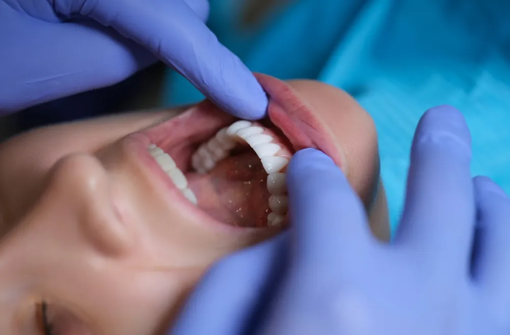
Small chips at the incisal edge sometimes allow for composite repair. The dentist roughens the ceramic surface. The dentist applies silane and bonding agent. Then the dentist adds composite resin to rebuild the missing fragment. This repair works best for tiny chips. It does not restore the original strength of the veneer. The repaired area remains a weak point. Patients must avoid biting forces on the repaired edge. Polishing can address minor surface scratches. The dentist uses fine diamond pastes to restore smoothness. This procedure improves esthetics and reduces plaque retention.
When Does a Veneer Need Replacement?
Replacement becomes necessary when the damage compromises function or esthetics. Large fractures, cracks extending across the veneer, or debonding that cannot be successfully recemented all require replacement. Secondary decay under the veneer also mandates replacement. The dentist must remove the old veneer. The dentist treats the decay. Then the dentist places a new veneer. Marginal staining with confirmed microleakage is another indication for replacement. Your dentist will evaluate the underlying tooth structure. If the tooth remains healthy, a new E.max veneer usually provides another decade or more of service.
What Happens During Veneer Replacement?
Veneer replacement follows a similar process to initial placement. The dentist removes the old veneer carefully. The dentist preserves as much enamel as possible. The dentist cleans the tooth surface. The dentist takes new impressions or digital scans. The lab fabricates a new veneer. The dentist bonds the new restoration. The entire process typically requires two visits. Some practices with in-house CAD/CAM systems can complete replacement in a single visit. After replacement, the patient follows the same care routine as with the original veneers.
How Can Patients Make E.max Veneers Last Longer?
Patients control much of their veneer destiny. The following habits stack the odds in favor of longevity.
Why Are Regular Checkups Essential?
Visit your dentist every six months. Your dentist examines the veneer margins. Your dentist checks for early signs of decay, debonding, or fracture. Your dentist evaluates your bite. Your dentist also performs professional cleaning. This cleaning removes calculus and stain. It keeps the gums healthy. Early intervention prevents small problems from becoming major failures. Rinke et al. (2020) emphasized that regular recall visits contributed to their high 10-year survival rates.
How Does a Night Guard Protect Veneers?
We have already discussed night guards for bruxism. But even patients without diagnosed bruxism can benefit from a night guard. Many people grind or clench occasionally during stressful periods. A night guard provides insurance against these episodes. It costs far less than replacing a veneer. Your dentist can fabricate a custom guard that fits comfortably. Wear it every night. Clean it daily. Replace it when it shows wear.
Why Do Healthy Gums Matter?
Healthy gums frame your veneers beautifully. They also seal the margin against bacterial invasion. Brush gently along the gumline. Use a soft brush. Do not scrub aggressively. Aggressive brushing causes gum recession. Floss carefully around each veneer. Do not snap the floss into the gum. Slide it gently under the contact point. If you notice bleeding, swelling, or tenderness, see your dentist. These symptoms indicate gingivitis. Treat gingivitis early. It is reversible. Untreated gingivitis becomes periodontitis. Periodontitis is not reversible. It destroys the foundation of your veneers.
Does Smoking Harm Veneers?
Smoking harms veneers in multiple ways. First, nicotine and tar stain the resin cement. They also stain the exposed tooth structure at the margin. These stains are difficult to remove. They create a visible line at the veneer edge. Second, smoking reduces blood flow to the gums. This reduction impairs the immune response. Smokers develop gum disease more readily. Gresnigt et al. (2019) found that smoking habits negatively affected the success rate of ceramic laminate veneers due to color changes and biological complications. Third, smoking slows healing after any dental procedure. If you want your E.max veneers to last, quit smoking. Your veneers, your gums, and your overall health will all benefit.
What Toothpaste Should Patients Use?
Use a non-abrasive fluoride toothpaste. Avoid whitening pastes with harsh abrasives. Avoid charcoal-based toothpastes. These products scratch the ceramic surface. They also scratch the exposed enamel at the margin. Over time, these scratches accumulate stains. They also create pathways for bacteria. Your dentist can recommend a suitable toothpaste brand. Many major manufacturers offer non-abrasive formulas specifically designed for cosmetic dental work.
Why Follow the Dentist's Care Instructions?
Your dentist knows your specific risk factors. Your dentist may recommend additional measures based on your bite, your habits, and your oral health status. Follow these instructions precisely. If your dentist recommends a specific mouthwash, use it. If your dentist suggests more frequent cleanings, schedule them. If your dentist advises dietary modifications, implement them. The dentist's recommendations are personalized. They address your unique situation. Ignoring professional advice is one of the fastest ways to shorten veneer lifespan.
Who Qualifies for Long-Lasting E.max Veneers?
Not every patient is an ideal candidate. Proper case selection sets the stage for decades of success.
Do Patients Need Healthy Teeth and Gums?
Yes. Ideal candidates have healthy teeth and gums. They have no active decay. They have no untreated periodontal disease. They have sufficient enamel for bonding. The 2024 substrate meta-analysis confirmed that enamel-bonded veneers achieve 99% survival. Patients with heavily restored teeth, severe wear, or extensive dentin exposure face higher risks. These patients may need crowns instead of veneers. Alternatively, they may need preliminary treatments like fillings or gum therapy before veneer placement.
Can Bruxism Patients Get E.max Veneers?
Bruxism patients can get E.max veneers, but they need special precautions. The dentist must design the veneers with reduced occlusal load. The dentist must provide a custom night guard before cementation. The patient must commit to wearing the guard every night. Faus-Matoses et al. (2020) showed that occlusal splints significantly improve survival in bruxism patients. Without a night guard, bruxism patients face substantially higher fracture and debonding rates. An honest dentist will discuss these risks openly. Some severe bruxers may be better candidates for zirconia or full-coverage crowns.
Does Oral Hygiene Commitment Matter?
Absolutely. Patients who neglect brushing and flossing should not get veneers. Veneers do not eliminate the need for oral hygiene. In fact, they demand meticulous care. The margin between veneer and tooth is a plaque trap. Patients who are unwilling to brush twice daily, floss daily, and attend regular cleanings will experience early failure. Good candidates understand that veneers are a privilege, not a shortcut. They treat their restorations with the same respect they give their natural teeth.
Is E.max Right for Conservative Cosmetic Treatment?
E.max excels in conservative cosmetic cases. Patients with mild discoloration, small chips, minor spacing issues, or slight misalignment make excellent candidates. E.max allows the dentist to correct these problems with minimal tooth reduction. Patients who want a dramatic transformation with maximum tooth preservation should ask their dentist about E.max. The material's combination of strength, esthetics, and conservative preparation makes it the gold standard for modern veneer dentistry.
What Do Patients Ask Most About E.max Veneers?
How long do E.max veneers typically last? E.max veneers typically last 10 to 15 years. Clinical studies report 10-year survival rates between 91.8% and 99.7%. With excellent care, many patients enjoy 20 years or more.
Can E.max veneers last 20 years? Yes, they can. No large-scale study has specifically tracked E.max veneers for 20 years. However, data from related ceramic veneers suggests that 20-year survival is achievable. Layton and Walton (2007) reported 91% survival at 15 years and 91% at 20 years for porcelain veneers. E.max's superior strength suggests comparable or better potential.
Do E.max veneers stain over time? The ceramic itself does not stain. E.max has excellent color stability. However, the resin cement at the margin can discolor. The exposed tooth structure can also stain. Regular cleanings and avoidance of staining foods and drinks minimize this issue.
Can teeth decay underneath E.max veneers? Yes. Decay can develop at the margin. It can also occur if the veneer debonds and bacteria seep underneath. Good oral hygiene and regular checkups prevent this problem.
Are E.max veneers stronger than porcelain veneers? E.max is a type of porcelain veneer. It is stronger than traditional feldspathic porcelain. E.max has a flexural strength of 360 to 400 MPa. Feldspathic porcelain has a flexural strength around 154 MPa. E.max resists fracture better than older porcelain types.
Can E.max veneers chip? Yes, they can chip. The risk is low but real. Bruxism, biting hard objects, and trauma cause most chips. A night guard and dietary awareness reduce this risk dramatically.
Do E.max veneers require special toothpaste? They require non-abrasive toothpaste. Avoid charcoal, baking soda, and heavy whitening pastes. These products scratch the surface. Your dentist can recommend a suitable brand.
How often should E.max veneers be professionally checked? Every six months. Your dentist examines the margins, the bite, and the gum health. Early detection of problems prevents major failures.
Can grinding teeth shorten the lifespan of E.max veneers? Yes. Grinding applies destructive lateral forces. It is one of the leading causes of veneer fracture. A night guard is essential for grinders.
What is the most common reason E.max veneers fail? Fracture is the most common reason. It accounts for approximately 4% of failures over 10 years. Debonding ranks second at approximately 2%. Both are largely preventable with proper technique and patient habits.
What Is the Bottom Line on E.max Veneer Longevity?
E.max veneers represent one of the most reliable investments in cosmetic dentistry. They typically last 10 to 15 years. They often last 20 years or more. Clinical studies consistently report 10-year survival rates above 90%. Some studies report rates as high as 99.7%.
Your veneers will last longer if you choose an experienced dentist. The dentist must perform conservative enamel preparation. The dentist must use meticulous adhesive cementation. The dental lab must fabricate the veneer with precision. You must maintain excellent oral hygiene. You must wear a night guard if you grind your teeth. You must avoid using your teeth as tools. You must attend regular dental checkups.
E.max veneers are not indestructible. They are not permanent. But they are durable, beautiful, and predictable. View them as a long-term investment in your smile. Treat them with care. Partner with a skilled dental team. Do this, and your E.max veneers will reward you with decades of confident smiles.
References
AlJazairy, Yazeed H. "Survival Rates for Porcelain Laminate Veneers." European Journal of Prosthodontics and Restorative Dentistry, vol. 28, no. 4, 2020, pp. 360–368.
Al-Thobity, Ahmad M., and Omar Alsalman. "Flexural Properties of Three Lithium Disilicate Materials." Journal of King Saud University - Science, vol. 33, no. 8, 2021, PMC8589621.
Aslan, Yasemin U., et al. "Retrospective Analysis of Lithium Disilicate Laminate Veneers Applied by Experienced Dentists: 10-Year Results." International Journal of Prosthodontics, vol. 32, no. 5, 2019, pp. 471–473.
Demirekin, Zeynep, et al. "Clinical Performance of Pressable Glass-Ceramic Veneers after 5, 10, 15, and 20 Years: A Retrospective Case Series Study." Journal of Esthetic and Restorative Dentistry, vol. 31, no. 5, 2022, pp. 415–422.
Faus-Matoses, Vicente, et al. "An 8-Year Prospective Clinical Investigation on the Survival Rate of Feldspathic Veneers: Influence of Occlusal Splint in Patients with Bruxism." Journal of Dentistry, vol. 99, 2020, 103352.
Fotiadou, Christina, et al. "Survival and Success Rates of Lithium Disilicate Indirect Restorations." Journal of Dentistry, vol. 113, 2021, 103773.
Fradeani, Mauro, et al. "Porcelain Laminate Veneers: 6- to 12-Year Clinical Evaluation - A Retrospective Study." International Journal of Periodontics and Restorative Dentistry, vol. 25, no. 1, 2005, pp. 9–17.
Gonzalez-Martin, Oscar, et al. "Total Fracture Occurrence in Veneers: A Clinical Study." Journal of Prosthetic Dentistry, vol. 126, no. 4, 2021, pp. 531–537.
Gresnigt, Marco M. M., et al. "Randomized Clinical Trial on Indirect Resin Composite and Ceramic Laminate Veneers: Up to 10-Year Findings." Journal of Dentistry, vol. 86, 2019, pp. 102–109.
Imburgia, Mario, et al. "A Retrospective Clinical Study on 1075 Lithium Disilicate CAD/CAM Veneers with Feather-Edge Margins Cemented on 105 Patients." Journal of Esthetic and Restorative Dentistry, vol. 33, no. 5, 2021, pp. 710–719.
Klein, Pascal, et al. "Survival and Complication Rates of Feldspathic, Leucite-Reinforced, Lithium Disilicate and Zirconia Ceramic Laminate Veneers: A Systematic Review and Meta-Analysis." Journal of Dentistry, vol. 109, 2021, 103666.
Layton, Danielle M., and Terry R. Walton. "An Up to 16-Year Prospective Study of 304 Porcelain Veneers." International Journal of Prosthodontics, vol. 20, no. 4, 2007, pp. 389–396.
Malchiodi, Luca, et al. "Clinical and Esthetical Evaluation of 79 Lithium Disilicate Multilayered Anterior Veneers with a Medium Follow-Up of 3 Years." European Journal of Dentistry, vol. 13, no. 4, 2019, pp. 581–588.
Mazzetti, Thais, et al. "10-Year Practice-Based Evaluation of Ceramic and Direct Composite Veneers." Dental Materials, vol. 38, no. 5, 2022, pp. 898–906.
Morimoto, Susana, et al. "Main Clinical Outcomes of Feldspathic Porcelain and Glass-Ceramic Laminate Veneers: A Systematic Review and Meta-Analysis of Survival and Complication Rates." International Journal of Prosthodontics, vol. 29, no. 1, 2016, pp. 38–49.
Nejatidanesh, Farahnaz, et al. "Five-Year Clinical Outcomes and Survival of Chairside CAD/CAM Ceramic Laminate Veneers - A Retrospective Study." Journal of Prosthodontics, vol. 27, no. 4, 2018, pp. 338–344.
Rinke, Sven, et al. "10-Year Survival Rate of Ceramic Veneers in a Clinical Practice." Journal of Prosthetic Dentistry, vol. 124, no. 3, 2020, pp. 291–297.
Sen, Nazmiye, et al. "Retrospective Evaluation of Factors Affecting Long-Term Clinical Performance of CAD/CAM Laminate Veneers." International Journal of Prosthodontics, vol. 37, no. 2, 2024, pp. 215–223.
Willard, Andrew. "The Science and Application of IPS E.max Dental Ceramic." Journal of the American Dental Association, vol. 149, no. 1, 2018, pp. 14–17.
Zulkiffli, Sofya, et al. "Evaluation and Comparison of Mechanical Properties of Lithium Disilicate-Based CAD/CAM Blocks." Sains Malaysiana, vol. 53, no. 1, 2024, pp. 177–186.