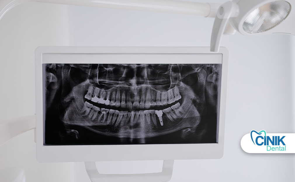
Panoramic dental X-rays provide a single, wide-view image of the entire mouth, including teeth, jaws, and surrounding bone structures. They are commonly used in orthodontics, oral surgery, and complex diagnostic cases to assess tooth position, jaw anatomy, wisdom teeth, and hidden pathologies such as cysts or tumors. The procedure is quick, painless, noninvasive, and uses a relatively low radiation dose compared to traditional X-rays.
Although panoramic X-rays are not required for routine dental check-ups, they are valuable when broader clinical insight is needed, such as for treatment planning, implant assessment, or evaluating abnormalities. When used appropriately, they offer comprehensive diagnostic information while balancing patient safety and clinical necessity.
1. What is a Panoramic Dental X-Ray
Quick Answer: A panoramic dental X-ray is a single image of the entire mouth taken by a rotating X-ray unit, used to assess teeth, jaws, and bone structure with a low radiation dose.
A panoramic dental X-ray is a broad view capable of capturing the whole mouth in a single image. A patient’s head remains still while a rotating X-ray unit moves around them, allowing dentists to examine bone structure, teeth position, and abnormalities. Such radiographs are important in orthodontics, oral surgery, and when traumatic injuries may require extensive examination. Simultaneously, the lower dose of radiation and ease of obtaining images suit growing children with multiple teeth changing (Puricelli, 2009).
2. Why Dentists Take Panoramic X-Rays
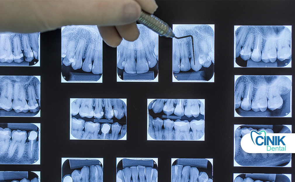
Quick Answer: Dentists take panoramic X-rays to view the entire jaw in one image, allowing assessment of tooth position, bone structure, dental development, hidden lesions, and unerupted teeth such as wisdom teeth.
Panoramic dental X-rays display the entire jaw in a single image and are employed to determine the position of teeth, bone structure, and potential abnormalities. Dentists take panoramic X-rays for several reasons. First, they allow assessment of bone anatomy and pathology, dental development, and the extent of dental treatment. Second, they evaluate the mandible and maxillofacial region for certain lesions, cysts, and tumours not visible through intraoral films. Third, they detect gross caries, periodontal disease, or other pathology overlooked on intraoral films. Lastly, they are valuable for identifying unerupted teeth, especially wisdom teeth, and establishing their relationship to adjacent teeth (Ozmen and Basak Ayna, 2024).
3. Is a Panoramic X-Ray Always Necessary
Quick Answer: No. Panoramic X-rays are not needed for routine check-ups and are used only when specific clinical indications exist, such as suspected abnormalities, orthodontic planning, implant assessment, or wisdom tooth evaluation.
Most of the time, however, a panoramic x-ray is not necessary. For the typical check-up, for instance, full records may include a bite-wing survey that monitors for cavities in the posterior teeth. Only if there’s a clinical indication suggesting a need to examine the anterior teeth, such as a missing tooth, would a dentist request a panoramic x-ray (Couto Ramos et al., 2016). In other situations a panoramic x-ray might be helpful but not strictly required:
Signs of a cyst, tumor, or other abnormality detected in a periapical radiograph. Ongoing orthodontic treatment.
Assessment of cases involving pre-existing dental implants.
Evaluation of wisdom tooth extraction cases already treated elsewhere.
Conditions of extended treatment requiring far more than bite-wing radiographs.
In these cases the selection of a panoramic x-ray must balance risks, benefits, and alternatives. All conditions requiring dental treatment, but no clinical or radiographic indication suggesting a need for a particular radiograph, are also instances when a panoramic x-ray remains nonessential (Ozmen and Basak Ayna, 2024).
4. How a Panoramic Dental X-Ray Is Taken
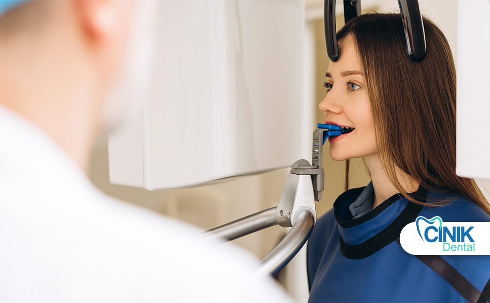
Quick Answer: A panoramic dental X-ray is taken by rotating an X-ray source and detector around the patient’s head, capturing a single image of the entire jaw in about 12–24 seconds with a low radiation dose.
Panoramic radiographs are a simple and widely used technique in dental specialties that involve synchronized rotation of an X-ray source and a stationary image receptor around the patient's head. This imaging method captures the entire maxillomandibular structure, including dental arches and related structures. It is beneficial because it involves a lower radiation dose, costs less, and images a larger area compared to intraoral radiographs. Panoramic radiography is recommended for patients with transitional or permanent dentition and can evaluate teeth, bone lesions, fractures, and foreign bodies (Couto Ramos et al., 2016).
A panoramic dental X-ray machine consists of a circular tube that emits the X-ray beam and a flat rectangular plate that receives the X-ray projection. The patient is positioned between the tube and the plate so that the X-ray beam is parallel to the plane of the teeth and the flat plate remains perpendicular to the horizontal plane passing through the center of the condyles. At the beginning of the exposure, the tube and the plate are positioned at an angle adjacent to the midline of the patient’s skull. The movement of the tube is diverging, while the plate moves in a horizontal straight line along the midline. At the completion of the action, the tube and the plate are positioned at the same angles but on opposite sides of the skull. Since the tube rotation occurs synchronously, while the tube and the plate are moving, a series of overlapping X-ray images are taken; the whole process lasts between 12 and 24 seconds.
5. Does a Panoramic X-Ray Hurt
Quick Answer: No. A panoramic X-ray is painless, noninvasive, and involves only a brief sensation of mild warmth or tingling.
Dental X-ray surveys help dentists make decisions about patients’ oral health, treatments, and hygiene. Panoramic X-rays or panoramic, generically exposed images allow a single depiction of the maxillary complex. They help outline the dentition contour, bone structure, and teeth alignment in both the upper and lower jaw (眞之, 2018). These X-rays expose the patient to significantly lower radiation levels than conventional periapical X-ray techniques.
The patient only briefly experiences a little tingling or heat over the neck or below the jaw. A panoramic X-ray does not hurt and does not pose concerning problems for the patient.
6. Conclusion
The article explained panoramic dental X-rays, large images that capture the entire jaw’s teeth position, surrounding bone structure, sinuses, and possible tumors, cysts, or infections. A dentist may recommend one to collect comprehensive information about a patient’s dental health, especially when planning orthodontic, surgical, or wisdom tooth removal procedures. The X-ray uses a minimal dose of radiation and is noninvasive and painless.
Panoramic dental X-rays are not required during every dental examination, but they can provide valuable information about jaw growth, wisdom teeth, and tooth position eruption problems. Cinematic panoramic X-rays are even more detailed, delivering true-to-life images that can be interpreted in motion.
References:
Puricelli, E. "Panorametry: suggestion of a method for mandibular measurements on panoramic radiographs." 2009. ncbi.nlm.nih.gov
Ozmen, B. and Basak Ayna, N. "Occlusal Radiographs in a Turkish Pediatric Subpopulation." 2024. ncbi.nlm.nih.gov
Couto Ramos, B., Raquel da Silva Izar, B., Lourdes Costa Pereira, J., Sena Souza, P., Scigliano Valerio, C., Mesquita Tuji, F., and Ricardo Manzi, F. "Formation of ghost images due to metal objects on the surface of the patient's face: A pictorial essay." 2016. ncbi.nlm.nih.gov
眞之, 牧 "Generation of a 2.5D Dental Panoramic Image." 2018. [PDF]


