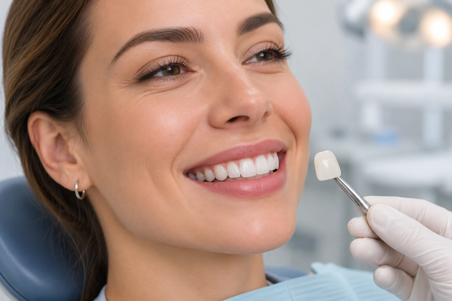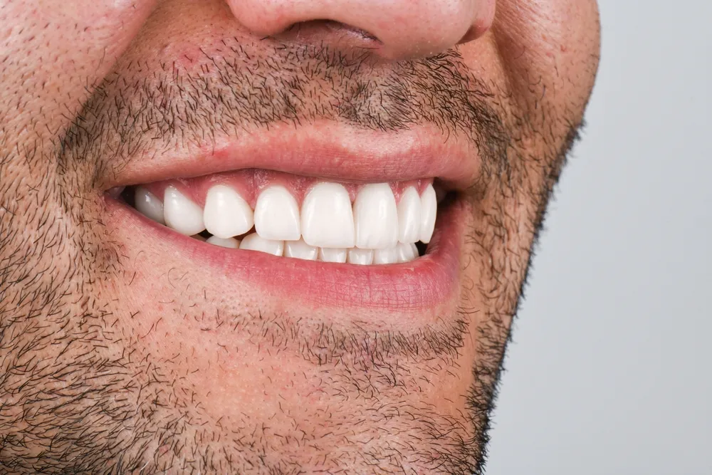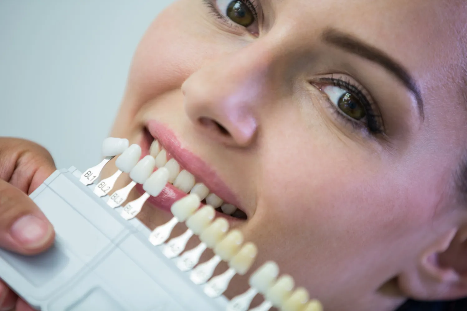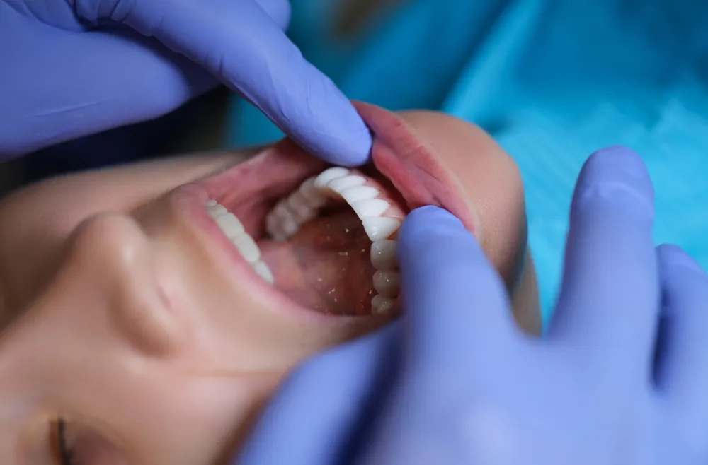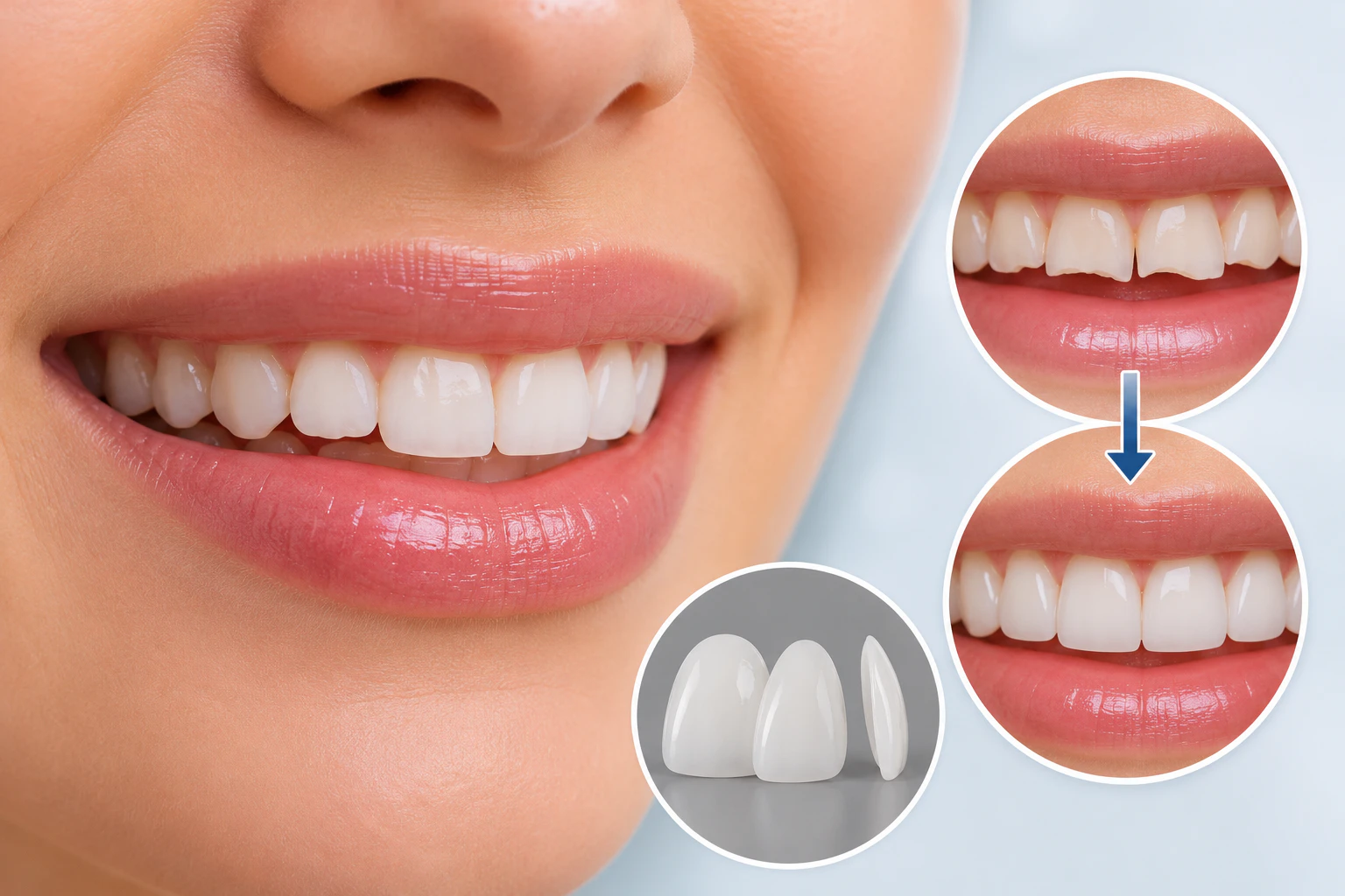
E.max veneers are custom-made ceramic shells that dentists bond to the front surfaces of chipped, shortened, or worn teeth to restore their original contour and appearance. Manufacturers produce them from lithium disilicate glass-ceramic, a material that combines high flexural strength with enamel-like translucency. Because of these two properties, clinicians widely use E.max veneers for anterior restorations that must look natural and withstand daily function.
A veneer works as a thin facing, usually 0.3 to 0.7 millimeters thick. The dentist prepares a controlled layer of enamel, records the tooth shape, and bonds the finished ceramic shell with resin cement. The restoration then recreates the visible portion of the tooth, including the biting edge and the light-reflecting surface texture. Magne and Belser describe this approach as a biomimetic one, because the bonded ceramic reproduces the mechanical and optical behavior of natural enamel (Magne and Belser 2002).
E.max veneers differ from conventional feldspathic porcelain veneers and composite veneers in both composition and performance. Feldspathic porcelain offers excellent aesthetics but lower strength. Composite resin costs less and allows chairside repair, yet it stains and loses surface polish faster. Lithium disilicate sits between these options: it delivers strong aesthetics, and laboratory data report flexural strength around 360 to 400 megapascals (Guess et al. 2011). Dentists apply these restorations to chipped enamel, worn incisal edges, uneven tooth lengths, and discoloration that accompanies surface damage.
How an E.max veneer is placed
A thin ceramic shell, usually 0.3 to 0.7 mm thick, is bonded to the front surface of the tooth.
How Do E.max Veneers Repair Chipped Teeth?
E.max veneers repair chipped teeth by covering the damaged front surface with a ceramic shell that recreates the missing portion of the tooth. This solution suits minor and moderate enamel damage, where the chip affects appearance but leaves enough healthy structure for bonding.
When a front tooth loses a corner or a section of its edge, the dentist first maps the defect and the surrounding enamel. The laboratory then fabricates a veneer that rebuilds the symmetrical incisal edge and restores the natural line angles of the tooth. The translucency of lithium disilicate plays a central role here. Light passes through the ceramic and reflects from the underlying tooth, so the restoration blends with neighboring teeth instead of looking opaque or flat (Guess et al. 2011).
Case selection determines success. A superficial enamel chip responds well to a veneer. A fracture that reaches the dentin or the pulp requires a different plan, because deeper damage changes the biological and mechanical status of the tooth. The next sections explain how clinicians make that distinction and why it matters for long-term outcomes.
Can E.max Veneers Restore Teeth Worn Down by Grinding?
Yes, E.max veneers can restore the length and appearance of anterior teeth worn down by grinding, provided the patient still has adequate enamel and the dentist addresses the grinding habit itself. Bruxism gradually shortens teeth and flattens their biting edges, a process called attrition. Population studies estimate that awake and sleep bruxism affect a substantial share of adults, with reported prevalence ranging roughly from 8 to 31 percent depending on the diagnostic method (Manfredini et al. 2013).
Restoring worn teeth without controlling the cause invites failure. Repeated clenching and grinding generate forces far beyond normal chewing, and these forces can chip the ceramic or break the adhesive bond. Dentists therefore evaluate enamel thickness, tooth structure, and the occlusion before treatment. After bonding the veneers, they usually prescribe a protective night guard to shield the restorations during sleep. This combined approach, restoration plus bite management, forms the standard of care for bruxism-related wear.
What Types of Tooth Damage Can E.max Veneers Correct?
E.max veneers correct surface-level and contour-level damage of front teeth, not deep structural fractures. The following list summarizes the conditions that respond well to ceramic veneers:
Minor enamel chips and irregular tooth edges
Superficial cracks and visible enamel imperfections
Attrition caused by grinding or clenching
Erosion-related changes in tooth shape and surface texture
Uneven incisal edges and shortened front teeth
Discoloration that accompanies structural surface defects
Each condition on this list shares one feature: the damage stays within the enamel or the outer dentin, and the tooth retains its structural integrity. Once damage crosses that boundary, the treatment logic changes, and the dentist must consider full-coverage options instead.
Who Is a Suitable Candidate for E.max Veneers?
A suitable candidate has enough healthy enamel for predictable bonding, healthy gums, and a stable or manageable bite. Adhesive dentistry depends on enamel, because resin cement bonds to enamel far more reliably than to dentin. Research on preparation design confirms that conservative preparations within enamel produce the most durable results (Edelhoff and Sorensen 2002).
Typical candidates include patients with chipped or worn front teeth but adequate remaining structure, individuals with healthy periodontal tissues and no untreated infections, and people who want to improve both the appearance and the proportions of their smile. Patients with stable occlusion also qualify, as do some grinders who accept night guard therapy. A clinical examination remains essential before confirming eligibility, because photographs and descriptions cannot reveal enamel thickness, hidden cracks, or bite problems.
When Are E.max Veneers Not the Right Treatment?
E.max veneers are not the right treatment when a tooth has lost its structural integrity or when the mouth presents uncontrolled disease or force problems. Contraindications include:
Extensive fractures that compromise the load-bearing structure of the tooth
Damage extending below the gumline or involving the root
Severe tooth decay or active periodontal disease
Insufficient enamel for reliable adhesive bonding
Uncontrolled bruxism or significant bite abnormalities
Pulp damage that requires root canal treatment before any cosmetic restoration
In these situations, the dentist treats the disease first and then chooses a restoration that protects the whole tooth, such as a crown. Recognizing these limits protects patients from premature failure, and it leads directly to the clinical decision discussed next.
How Do Dentists Decide Between a Veneer and a Crown?
Veneer or crown? It depends on how deep the damage goes
Select a type of damage to see how dentists usually approach it.
A minor chip that stays within the enamel
The veneer rebuilds the missing edge or corner and restores the natural line angles. Enough healthy enamel remains for a reliable bond.
Fine surface lines within the enamel
These are usually cosmetic and a veneer can cover them. The dentist first confirms how deep the crack runs, using X-rays and transillumination.
Damage that reaches the dentin
A deeper fracture weakens the load-bearing structure of the tooth. A crown encircles the whole tooth and protects it better than a front-surface veneer.
Damage involving the pulp or root
The pulp needs root canal treatment before any cosmetic restoration, typically followed by a crown. Vertical cracks running toward the root may require a crown or even extraction.
Illustration for general information only. The final decision requires a clinical examination and dental imaging.
Dentists decide between a veneer and a crown by measuring how much healthy tooth structure remains and where the damage sits. A veneer covers only the front surface, so it works when damage is cosmetic and the tooth body stays strong. A crown encircles the entire tooth, so it suits teeth weakened by large fractures, extensive decay, or root canal treatment.
Fracture depth, location, and direction guide this judgment. A horizontal chip at the biting edge often accepts a veneer. A vertical crack running toward the root often demands a crown or even extraction. Clinical examination and dental imaging, including radiographs and transillumination, supply the evidence for this choice. Systematic reviews show that both options perform well when matched to the right indication: lithium disilicate single crowns achieve survival rates above 96 percent at five years, while glass-ceramic veneers show similarly high survival when bonded to enamel (Pieger, Salman, and Bidra 2014; Morimoto et al. 2016).
E.max Veneers vs Composite Bonding for Chipped Teeth: Which Option Fits Better?
E.max veneers fit patients who need a comprehensive and durable cosmetic correction, while composite bonding fits very small chips that require a fast, reversible repair. The two materials differ in application, appearance, maintenance, and the amount of tooth preparation involved. The following table compares both options with dental crowns, the third common alternative for damaged front teeth.
Criterion | E.max Veneers | Composite Bonding | Dental Crowns |
Material | Lithium disilicate glass-ceramic | Composite resin | Ceramic or porcelain-fused materials |
Tooth coverage | Front surface and edge | Localized defect only | Entire tooth |
Tooth preparation | Conservative, usually 0.3 to 0.7 mm of enamel | Minimal or none | Substantial, all surfaces |
Stain resistance | High | Moderate, stains over time | High |
Surface polish retention | Long-lasting | Degrades within years | Long-lasting |
Repairability | Limited, usually requires replacement | Easy chairside repair | Limited |
Typical longevity | 10 years and beyond in most studies | 3 to 7 years on average | 10 to 15 years |
Best indication | Cosmetic damage with adequate enamel | Very small chips, budget-limited cases | Structurally weakened teeth |
Veneers, bonding and crowns at a glance
Typical ranges from the comparison table above. Individual results depend on the case.
Typical longevity (years)
How much natural tooth is removed
Bar lengths for tooth reduction are illustrative, not to scale.
Composite resin bonds directly to the tooth in one visit, and the dentist can polish or patch it after minor damage. Ceramic cannot be patched reliably, but it holds its color and gloss far longer. For a single tiny chip, bonding often wins. For multiple worn or discolored teeth, E.max veneers usually deliver the more complete and stable result (Fradeani, Redemagni, and Corrado 2005).
Why Is Lithium Disilicate Used for E.max Veneers?
Manufacturers use lithium disilicate for E.max veneers because the material combines mechanical strength with optical behavior close to natural enamel. Lithium disilicate is a glass-ceramic: needle-shaped lithium disilicate crystals grow inside a glassy matrix, and these interlocking crystals deflect cracks and raise the fracture resistance of the material. Laboratory measurements place its flexural strength around 360 to 400 megapascals, several times higher than conventional feldspathic porcelain (Guess et al. 2011).
The glassy phase gives the ceramic its translucency. Technicians can adjust opacity, shade, and surface characterization, so the restoration reproduces the depth and light transmission of natural enamel. Ceramic thickness and restoration design also shape the final optical result: a thicker veneer masks discoloration better, while a thinner one preserves more tooth and transmits more underlying color.
Strength alone does not guarantee success. The resin cement bond carries much of the load, and laboratory fabrication quality controls the fit of the margins. Clinical research confirms that lithium disilicate restorations bonded to enamel with proper adhesive protocols achieve high survival rates, while the same material can still chip or fracture under excessive force (Beier et al. 2012). Material performance and clinical handling therefore work as one system.
What Happens During the E.max Veneer Treatment Process?
The E.max veneer treatment process moves through eight clinical steps, from diagnosis to final bite adjustment. Each step builds on the previous one, so precision at the start determines the quality of the result.
The E.max veneer treatment, step by step
Each step builds on the previous one, so precision at the start shapes the final result.
- Step 1
Initial examination
Tooth damage, enamel, gums and bite are evaluated. X-rays and photos reveal hidden cracks or decay.
- Step 2
Digital smile planning
Proportions, shade and final shape are designed. You preview the planned outcome before any enamel is touched.
- Step 3
Tooth preparation
A controlled layer of enamel, usually 0.3 to 0.7 mm, is removed to make space for the ceramic.
- Step 4
Impressions or digital scan
The exact tooth dimensions are recorded so the laboratory can make veneers with tight, precise margins.
- Step 5
Temporary veneers
They protect prepared teeth and keep your appearance in the meantime. Minimal-preparation cases may skip this step.
- Step 6
Try-in appointment
Fit, color, shape and alignment are checked with a temporary paste. You approve the look before bonding.
- Step 7
Permanent bonding
The ceramic and enamel are conditioned and each veneer is seated with light-cured resin cement.
- Step 8
Final bite assessment
Bite contacts are checked and adjusted to reduce stress on the ceramic edges.
What Happens During the Initial Examination?
The dentist evaluates the tooth damage, enamel condition, gum health, and occlusion. Radiographs and photographs document the baseline state and reveal hidden problems such as cracks or decay.
What Does Digital Smile Planning Involve?
The team assesses tooth proportions, selects the target shade, and designs the final restoration shape. Digital previews let the patient review the planned outcome before any enamel removal.
How Does Tooth Preparation Work?
The dentist removes a controlled layer of enamel, usually 0.3 to 0.7 millimeters, to create space for the ceramic. Measured studies of preparation designs show that conservative veneer preparations remove far less tissue than crown preparations (Edelhoff and Sorensen 2002).
Why Are Impressions or Digital Scans Taken?
An impression or an intraoral scan records the exact tooth dimensions. The dental laboratory uses this record to fabricate veneers that fit the prepared surfaces with tight margins.
When Are Temporary Veneers Necessary?
Temporary veneers protect prepared teeth and maintain appearance when the dentist removes a visible amount of enamel. Minimal-preparation cases sometimes skip this step.
What Is the Purpose of the Try-In Appointment?
The dentist places the finished ceramics with a water-soluble try-in paste and checks fit, color, shape, and alignment. The patient approves the aesthetics before permanent bonding.
How Does Permanent Bonding Work?
The dentist etches the ceramic with hydrofluoric acid, applies silane, conditions the enamel, and seats each veneer with light-cured resin cement. Adhesive protocols of this type produce the durable enamel bond documented in long-term clinical studies (Cortellini and Canale 2012).
What Happens at the Final Bite Assessment?
The dentist checks occlusal contacts in centric and eccentric movements and adjusts any premature contact. This step reduces stress concentration on the ceramic edges.
How Long Do E.max Veneers Last on Chipped or Worn Teeth?
Clinical studies report that glass-ceramic veneers commonly survive 10 years and longer when dentists bond them to enamel and select cases carefully. A systematic review and meta-analysis found estimated survival rates above 90 percent at 10 years for laminate veneers, with enamel bonding identified as a key success factor (Morimoto et al. 2016). A longitudinal study of 323 porcelain veneers reached similar conclusions over observation periods of up to 11 years (Granell-Ruíz et al. 2014).
Several factors move an individual case above or below these averages. Enamel preservation matters most, because bonds to dentin degrade faster. Chewing habits and excessive biting forces, such as biting hard objects, concentrate stress at the ceramic margins. Untreated bruxism and bite problems accelerate chipping and debonding. Regular dental examinations help the clinician detect early signs of wear, margin leakage, or debonding before a small defect becomes a failure.
Longevity does not mean permanence. A veneer that functions for 15 years may still need replacement because of material fatigue, gum recession, or changes in the underlying tooth. Patients should view veneers as long-term restorations with a finite service life, not as lifetime devices.
How Should You Care for E.max Veneers After Treatment?
You care for E.max veneers with the same daily hygiene as natural teeth, plus a few protective habits that shield the ceramic from mechanical stress. Consistent aftercare protects both the restoration and the tooth underneath it.
Brush twice daily with a soft-bristled toothbrush and non-abrasive toothpaste.
Clean between the teeth every day with floss or interdental brushes.
Avoid biting hard objects such as ice, pens, fingernails, and hard candies.
Wear the dentist-prescribed night guard if you grind your teeth.
Attend routine dental examinations and professional cleanings.
Never use your teeth as tools to open packaging or tear materials.
Remember that veneers cannot be whitened; bleaching agents do not change ceramic color.
These habits address the main failure modes of ceramic veneers: marginal decay, surface wear of the opposing teeth, and mechanical overload. The next section explains what happens when these defenses fail.
What Are the Possible Risks of E.max Veneers for Worn Teeth?
The possible risks of E.max veneers include sensitivity, ceramic fracture, debonding, gum irritation, secondary decay, and the irreversible loss of prepared enamel. Clinical failure analyses identify fracture and debonding as the most frequent technical complications of all-ceramic restorations (Beier et al. 2012).
Post-treatment sensitivity can follow enamel preparation, especially when preparation reaches dentin. Ceramic chipping occurs under excessive occlusal pressure or impact. Debonding results from inadequate bonding conditions, contamination during cementation, or sustained mechanical stress. Poorly designed restoration margins irritate the gums and complicate cleaning, which raises the risk of secondary decay around exposed margins. Finally, conventional preparation removes enamel permanently, so the tooth will always require a restoration afterward.
Appropriate case selection and disciplined treatment planning control most of these risks. This is why the contraindications listed earlier, such as uncontrolled bruxism or insufficient enamel, carry so much weight in the diagnostic phase.
How Much Do E.max Veneers Cost for Chipped or Worn Teeth?
The cost of E.max veneers varies widely, because the price reflects the number of teeth, the laboratory, the clinician, and any supporting procedures. No single figure applies to every patient, so an individualized assessment is the only reliable source of pricing.
Several factors drive the final investment. Each additional tooth adds laboratory and chair time. High-quality laboratories with master technicians charge more, and their fee reflects customization of shade, texture, and translucency. Preparatory procedures such as gum contouring, treatment of decay, or bite correction raise the total. Geographic location and clinician experience also influence fees.
A long-term view helps the comparison with composite bonding. Bonding costs less upfront but typically needs polishing, repair, or replacement within several years. Ceramic veneers cost more at the start and hold their appearance longer, which many patients find favorable over a decade of use. A written treatment plan after clinical examination gives the most accurate picture for an individual case.
Frequently Asked Questions About E.max Veneers for Chipped or Worn Teeth
This section answers the questions patients ask most often before choosing E.max veneers for chipped or worn teeth. Each answer summarizes the evidence discussed in the sections above.
Can E.max Veneers Fix a Chipped Front Tooth?
Yes, E.max veneers can fix a chipped front tooth when the damage stays within the enamel and enough healthy structure remains for bonding. A minor cosmetic chip accepts a veneer predictably. A deeper fracture that reaches the dentin or pulp needs a different restoration, such as a crown, after the dentist completes any necessary underlying treatment.
Can E.max Veneers Restore Teeth Shortened by Grinding?
Yes, veneers can rebuild the length and appearance of teeth shortened by grinding. The critical condition is control of the bruxism itself. Without a night guard and bite management, the same forces that wore the natural enamel will attack the ceramic.
Are E.max Veneers Strong Enough for Worn Teeth?
E.max veneers are strong enough for worn anterior teeth when bonding, thickness, and occlusion are properly managed. Lithium disilicate offers flexural strength around 360 to 400 megapascals (Guess et al. 2011), and clinical reviews confirm high survival rates for glass-ceramic veneers bonded to enamel (Morimoto et al. 2016). Bite forces, veneer thickness, and adhesive quality together determine the margin of safety in each case.
Can E.max Veneers Be Placed Without Shaving Natural Teeth?
In selected cases, yes. No-preparation and minimal-preparation veneers exist alongside conventional designs. The existing tooth shape and the available space decide which design fits. Adding ceramic to an already prominent tooth without preparation would create bulk, so many cases still require a small amount of enamel reduction.
Are E.max Veneers Better Than Dental Bonding for Chipped Teeth?
Neither option is universally better; the extent of the damage determines the right choice. Bonding suits very small chips because it is fast, inexpensive, and repairable. E.max veneers suit broader cosmetic damage because ceramic resists staining, keeps its polish, and lasts longer. The comparison table earlier in this article summarizes the trade-offs.
Can E.max Veneers Break If I Grind My Teeth?
Yes, repeated clenching and grinding can chip or debond ceramic veneers. Bruxism generates forces well beyond normal chewing, and no restorative material tolerates unlimited load. A custom night guard worn during sleep protects the restorations and remains the standard protective measure for grinders with veneers.
Can E.max Veneers Cover Cracks in Natural Teeth?
E.max veneers can cover superficial enamel cracks, but dentists must first establish how deep the crack runs. Fine surface lines within enamel are usually cosmetic. Cracks that extend into dentin or toward the root change the prognosis of the tooth and may require a crown or other treatment instead.
Do E.max Veneers Require Replacement?
Yes, veneers can require replacement over time. Wear, chipping, debonding, gum recession, or decay at the margins may all lead to renewal of the restoration. Regular dental monitoring catches these changes early, and timely replacement protects the underlying tooth.
Conclusion: Are E.max Veneers a Good Solution for Chipped or Worn Teeth?
E.max veneers are a well-documented and conservative solution for chipped or worn front teeth, provided the damage stays cosmetic and the case selection is careful. Lithium disilicate ceramic restores tooth contour, length, and natural-looking translucency while preserving most of the healthy structure underneath. Long-term studies report survival rates above 90 percent at 10 years for glass-ceramic veneers bonded to enamel (Morimoto et al. 2016).
Not every chipped or worn tooth needs a veneer. Small isolated chips often respond to composite bonding, and structurally weakened teeth need crowns. The decisive factors are enamel quantity, occlusion, and overall dental health, all of which require a clinical examination. An individualized assessment by a qualified dentist remains the reliable path to the right treatment choice.
References
Beier, Ulrike S., et al. "Clinical Long-Term Evaluation and Failure Characteristics of 1,335 All-Ceramic Restorations." International Journal of Prosthodontics, vol. 25, no. 1, 2012, pp. 70-78.
Cortellini, Davide, and Angelo Canale. "Bonding Lithium Disilicate Ceramic to Feather-Edge Tooth Preparations: A Minimally Invasive Treatment Concept." Journal of Adhesive Dentistry, vol. 14, no. 1, 2012, pp. 7-10.
Edelhoff, Daniel, and John A. Sorensen. "Tooth Structure Removal Associated with Various Preparation Designs for Anterior Teeth." Journal of Prosthetic Dentistry, vol. 87, no. 5, 2002, pp. 503-509.
Fradeani, Mauro, Mauro Redemagni, and Massimo Corrado. "Porcelain Laminate Veneers: 6- to 12-Year Clinical Evaluation. A Retrospective Study." International Journal of Periodontics and Restorative Dentistry, vol. 25, no. 1, 2005, pp. 9-17.
Granell-Ruíz, Maria, et al. "A Clinical Longitudinal Study of 323 Porcelain Laminate Veneers: Period of Study from 3 to 11 Years." Medicina Oral, Patología Oral y Cirugía Bucal, vol. 19, no. 6, 2014, pp. e531-e537.
Guess, Petra C., et al. "All-Ceramic Systems: Laboratory and Clinical Performance." Dental Clinics of North America, vol. 55, no. 2, 2011, pp. 333-352.
Magne, Pascal, and Urs C. Belser. Bonded Porcelain Restorations in the Anterior Dentition: A Biomimetic Approach. Quintessence Publishing, 2002.
Manfredini, Daniele, et al. "Epidemiology of Bruxism in Adults: A Systematic Review of the Literature." Journal of Orofacial Pain, vol. 27, no. 2, 2013, pp. 99-110.
Morimoto, Susana, et al. "Main Clinical Outcomes of Feldspathic Porcelain and Glass-Ceramic Laminate Veneers: A Systematic Review and Meta-Analysis of Survival and Complication Rates." International Journal of Prosthodontics, vol. 29, no. 1, 2016, pp. 38-49.
Pieger, Sven, Anas Salman, and Avinash S. Bidra. "Clinical Outcomes of Lithium Disilicate Single Crowns and Partial Fixed Dental Prostheses: A Systematic Review." Journal of Prosthetic Dentistry, vol. 112, no. 1, 2014, pp. 22-30.